 Podit
Podit
Aşağıdaki çalışma materyali, ders kaydından elde edilen bilgilerle hazırlanmıştır.
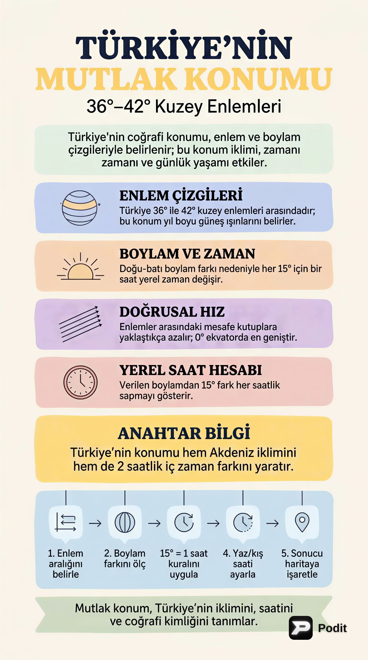
Türkiye'nin Mutlak Konumu ve Coğrafi Sonuçları
Bu çalışma materyali, bir yerin Dünya üzerindeki matematiksel konumunu ve bunun Türkiye üzerindeki etkilerini anlamanıza yardımcı olmak amacıyla hazırlanmıştır. Özellikle enlem ve boylam kavramları ile bunların coğrafi olaylar üzerindeki rolü üzerinde durulacaktır.
1. Mutlak Konum Nedir? 📚
Mutlak konum, bir yerin Dünya üzerindeki enlem ve boylam dereceleriyle ifade edilen matematiksel konumudur. Bu konum, o yerin iklim özelliklerinden yerel saatine, gece-gündüz sürelerinden mevsimlerin yaşanış biçimine kadar birçok coğrafi özelliğini doğrudan etkiler.
✅ Anahtar Bilgi: Mutlak konum, bir yerin "adresini" küresel koordinat sistemi üzerinde belirler.
2. Türkiye'nin Mutlak Konumu 🌍
Türkiye, Kuzey Yarım Küre'de ve başlangıç meridyeninin doğusunda yer alır. Coğrafi koordinatları şunlardır:
- Enlemler: 36° - 42° Kuzey Enlemleri arasında yer alır.
- Boylamlar: 26° - 45° Doğu Boylamları arasında yer alır.
Bu konum, Türkiye'nin coğrafi özelliklerini büyük ölçüde şekillendirir.
3. Enlemin Türkiye Üzerindeki Etkileri (36° - 42° Kuzey)
Enlem, Güneş ışınlarının geliş açısını ve dolayısıyla sıcaklık kuşaklarını, mevsimleri ve gece-gündüz sürelerini etkiler.
3.1. Mevsimlerin Oluşumu ve Gece-Gündüz Süreleri ☀️🌙
Türkiye'nin 36°-42° Kuzey enlemlerinde yer alması, Orta Kuşak'ta bulunmasını sağlar. Bu durumun sonuçları:
- Dört Mevsimin Belirgin Yaşanması: İlkbahar, yaz, sonbahar ve kış mevsimleri belirgin olarak yaşanır.
- Yaz-Kış Oluşumu: Güneş ışınlarının geliş açısı yıl içinde değiştiği için yaz ve kış mevsimleri oluşur.
- Gece-Gündüz Süreleri: Yıl içinde gece ve gündüz süreleri değişir. Kuzeye doğru gidildikçe (örneğin Antalya'dan Sinop'a) gece ile gündüz arasındaki süre farkı artar. Örneğin, yazın gündüzler daha uzun, kışın ise geceler daha uzundur.
3.2. Çizgisel Hız 💨
Dünya'nın kendi ekseni etrafındaki dönüş hızı (çizgisel hız), Ekvator'dan kutuplara doğru azalır.
- Türkiye'de güneyden kuzeye doğru gidildikçe (Ekvator'dan uzaklaşıldığı için) çizgisel hız azalır. Bu durum, Güneş'in doğuş ve batış sürelerini etkiler.
3.3. Gölge Boyları 📏
Güneş ışınlarının geliş açısı enleme göre değiştiği için, cisimlerin gölge boyları da enleme göre farklılık gösterir.
- Türkiye'de güneyden kuzeye gidildikçe Güneş ışınlarının geliş açısı küçüldüğü için gölge boyları uzar.
4. Boylamın Türkiye Üzerindeki Etkileri (26° - 45° Doğu)
Boylam, yerel saat farklılıklarının temel nedenidir. Dünya, kendi ekseni etrafında dönerken her meridyen Güneş'in karşısından farklı zamanlarda geçer.
4.1. Yerel Saat Farkları ⏰
- Güneş'in Konumu: Güneş, her boylam üzerinde farklı anlarda en tepe noktasına ulaşır. Bu da her boylamın kendine özgü bir yerel saate sahip olmasına neden olur.
- Doğu-Batı Zaman Farkı: Türkiye'nin en doğusu (45° Doğu boylamı) ile en batısı (26° Doğu boylamı) arasında 19 boylam farkı bulunur (45 - 26 = 19). Her boylam arası 4 dakika olduğu için, Türkiye'nin doğusu ile batısı arasında 19 x 4 = 76 dakikalık bir yerel saat farkı vardır.
- Ortak Saat Uygulaması: Ülke içinde saat birliğini sağlamak amacıyla ortak saat kullanılır. Türkiye, genellikle 45° Doğu boylamının (Iğdır'ın boylamı) yerel saatini ulusal saat olarak kullanır.
4.2. Güneş'in Doğuş ve Batış Saatleri 🌅
- Doğuda yer alan illerde Güneş daha erken doğar ve daha erken batar. Batıda yer alan illerde ise Güneş daha geç doğar ve daha geç batar.
5. Yerel Saat Hesaplamaları 💡
Yerel saat hesaplamaları, KPSS gibi sınavlarda sıkça karşılaşılan önemli bir konudur.
1️⃣ Boylam Farkını Bulma: İki yer arasındaki boylam farkı bulunur. 2️⃣ Zaman Farkını Hesaplama: Bulunan boylam farkı 4 dakika ile çarpılarak toplam zaman farkı bulunur. 3️⃣ Saati Belirleme: * Doğuya gidildikçe saat ileri alınır. * Batıya gidildikçe saat geri alınır.
Örnek: 30° Doğu boylamında saat 14:00 iken, 45° Doğu boylamında saat kaç olur?
- Boylam farkı: 45° - 30° = 15 boylam
- Zaman farkı: 15 boylam * 4 dakika/boylam = 60 dakika = 1 saat
- 45° Doğu, 30° Doğu'nun doğusunda olduğu için saat ileri alınır: 14:00 + 1 saat = 15:00.
6. Enlem ve Boylamlar Arasındaki Mesafeler 📏
Dünya üzerindeki enlem ve boylam çizgileri arasındaki mesafeler farklılık gösterir.
- Paraleller (Enlemler): Ardışık iki paralel (enlem) arasındaki mesafe her yerde yaklaşık olarak 111 kilometredir. Bu mesafe sabittir.
- Meridyenler (Boylamlar): Ardışık iki meridyen (boylam) arasındaki mesafe Ekvator üzerinde en geniştir (yaklaşık 111 km). Kutuplara doğru gidildikçe bu mesafe daralır ve meridyenler kutup noktalarında birleşir.
Bu bilgiler, Türkiye'nin mutlak konumunun coğrafi özelliklerini ve günlük yaşam üzerindeki etkilerini anlamanıza yardımcı olacaktır.