📚 Shoulder and Arm Musculature: A Comprehensive Study Guide
Source Information: This study material has been compiled from a combination of copy-pasted text (including anatomical figures and tables) and an audio lecture transcript. All information has been consolidated and presented in English for clarity and ease of study.
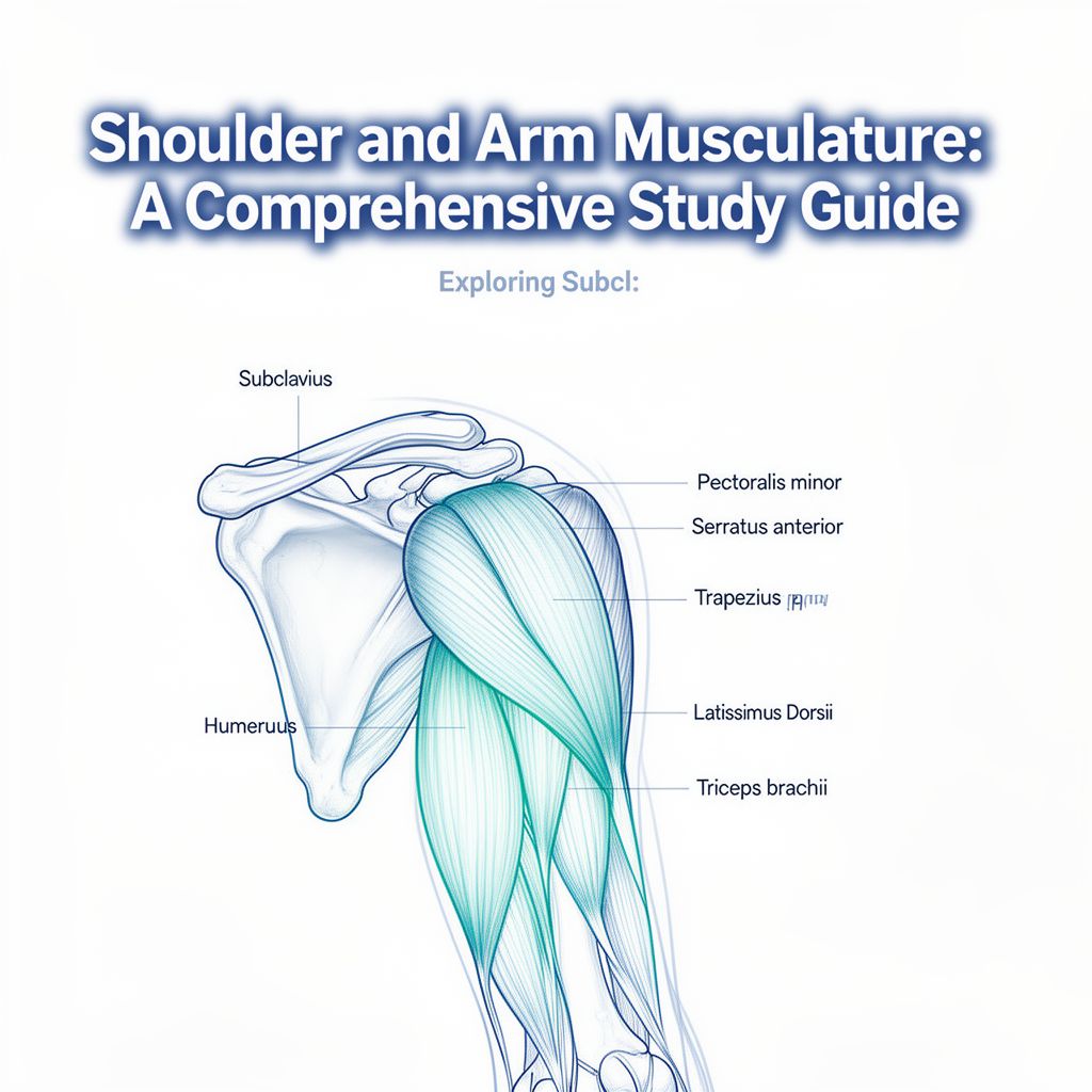
Introduction to Shoulder and Arm Musculature
This guide provides a detailed overview of the key muscles in the shoulder and arm regions. We will explore each muscle's origin, insertion, innervation, and primary actions, crucial for understanding human movement and anatomy.
1. Anterior Thoracic and Scapular Muscles
These muscles are located on the front of the chest and around the shoulder blade.
1.1. Subclavius
- Origin: 1st rib 🦴
- Insertion: Inferior surface of the clavicle
- Innervation: Nerve to subclavius (C5, C6)
- Action: ✅ Steadies the clavicle in the sternoclavicular joint.
1.2. Pectoralis Minor
- Origin: 3rd to 5th ribs 🦴
- Insertion: Coracoid process of the scapula
- Innervation: Medial pectoral nerve (C8, T1)
- Action:
- ✅ Draws the scapula downward.
- ✅ Causes the inferior angle of the scapula to move posteromedially.
- ✅ Rotates the glenoid cavity inferiorly.
- ✅ Assists in respiration.
1.3. Serratus Anterior
- Origin: 1st to 9th ribs 🦴
- Insertion: Costal and dorsal surfaces of the superior angle, medial border, and inferior angle of the scapula.
- Innervation: Long thoracic nerve (C5-C7)
- Action:
- Superior part: ✅ Lowers the raised arm.
- Entire muscle: ✅ Draws the scapula laterally forward.
- ✅ Elevates ribs when the shoulder is fixed.
- Inferior part: ✅ Rotates the inferior angle of the scapula laterally forward, enabling arm elevation above 90 degrees.
2. Posterior Thoracic and Scapular Muscles
These muscles are found on the upper back and around the shoulder blade.
2.1. Trapezius
- Origin: Occipital bone, nuchal ligament, and spinous processes of C1-T12 vertebrae 🦴
- Insertion: Lateral one-third of the clavicle, acromion, and scapular spine.
- Innervation: Accessory nerve (CN XI) and C3-C4 nerves of the cervical plexus.
- Action:
- Descending (Upper) part: ✅ Draws the scapula obliquely upward; rotates the glenoid cavity superiorly; tilts the head to the same side and rotates it to the opposite side.
- Transverse (Middle) part: ✅ Draws the scapula medially.
- Ascending (Lower) part: ✅ Draws the scapula medially downward.
- Entire muscle: ✅ Steadies the scapula on the thorax.
2.2. Levator Scapulae
- Origin: Transverse processes of C1-C4 vertebrae 🦴
- Insertion: Superior angle of the scapula.
- Innervation: Dorsal scapular nerve and cervical spinal nerves (C3-C4).
- Action: ✅ Draws the scapula medially upward while moving the inferior angle medially; inclines the neck to the same side.
2.3. Rhomboid Minor and Major
- Rhomboid Minor:
- Origin: Spinous processes of C6, C7 vertebrae 🦴
- Insertion: Medial border of the scapula (above the scapular spine).
- Rhomboid Major:
- Origin: Spinous processes of T1-T4 vertebrae 🦴
- Insertion: Medial border of the scapula (below the scapular spine).
- Innervation (both): Dorsal scapular nerve (C4-C5).
- Action (both): ✅ Steadies the scapula; draws the scapula medially upward.
3. Posterior Trunk and Arm Muscles
These include large muscles of the back and those connecting the trunk to the arm.
3.1. Latissimus Dorsi
- Origin: Spinous processes of T7-T12 vertebrae, thoracolumbar fascia, inferior angle of the scapula, 9th to 12th ribs, and posterior one-third of the iliac crest 🦴
- Insertion: Floor of the intertubercular groove of the humerus.
- Innervation: Thoracodorsal nerve (C6-C8).
- Action:
- ✅ Internal rotation of the arm.
- ✅ Adduction of the arm.
- ✅ Extension of the arm.
- ✅ Assists in respiration (often called the "cough muscle" 💡).
3.2. Teres Major
- Origin: Inferior angle of the scapula 🦴
- Insertion: Crest of the lesser tubercle of the humerus (anterior angle).
- Innervation: Lower subscapular nerve (C5, C6).
- Action:
- ✅ Internal rotation of the arm.
- ✅ Adduction of the arm.
- ✅ Extension of the arm.
4. Anterior Arm Muscles (Flexors)
These muscles are primarily responsible for flexing the elbow joint.
4.1. Biceps Brachii
- Origin:
- Long head: Supraglenoid tubercle of the scapula 🦴
- Short head: Coracoid process of the scapula 🦴
- Insertion: Radial tuberosity and bicipital aponeurosis.
- Innervation: Musculocutaneous nerve (C5-C6).
- Action:
- Elbow joint: ✅ Flexion; powerful supination (⚠️ especially when the elbow is flexed, as the lever arm is almost perpendicular to the axis of pronation/supination).
- Shoulder joint: ✅ Flexion; stabilization of the humeral head during deltoid contraction; abduction and internal rotation of the humerus.
4.2. Brachialis
- Origin: Distal half of the anterior surface of the humerus 🦴
- Insertion: Ulnar tuberosity.
- Innervation: Musculocutaneous nerve (C5-C6) and radial nerve (C7, minor contribution).
- Action: ✅ Flexion at the elbow joint.
5. Posterior Arm Muscles (Extensors)
These muscles are primarily responsible for extending the elbow joint.
5.1. Triceps Brachii
- Origin:
- Long head: Infraglenoid tubercle of the scapula 🦴
- Medial head: Posterior humerus, distal to the radial groove; medial intermuscular septum 🦴
- Lateral head: Posterior humerus, proximal to the radial groove; lateral intermuscular septum 🦴
- Insertion: Olecranon of the ulna.
- Innervation: Radial nerve (C6-C8).
- Action:
- Elbow joint: ✅ Extension.
- Shoulder joint (long head only): ✅ Extension and adduction.
5.2. Anconeus
- Origin: Lateral epicondyle of the humerus (sometimes from the posterior joint capsule) 🦴
- Insertion: Olecranon of the ulna (radial surface).
- Innervation: Radial nerve (C6-C8).
- Action: ✅ Extends the elbow and tightens its joint capsule.