The Visual Pathway, Reflexes, and Eye Movements - kapak
Bilim#visual pathway#optic nerve#lgb#visual cortex

The Visual Pathway, Reflexes, and Eye Movements

An academic overview of the visual pathway from retina to cortex, including visual field processing, magnocellular and parvocellular streams, light and accommodation reflexes, and conjugate eye movements.

juliamedicFebruary 22, 2026 ~25 dk toplam
01

Sesli Özet

9 dakika

Konuyu otobüste, koşarken, yolda dinleyerek öğren.

Sesli Özet

The Visual Pathway, Reflexes, and Eye Movements

0:008:39
02

Flash Kartlar

25 kart

Karta tıklayarak çevir. ← → ile gez, ⎵ ile çevir.

1 / 25
Tüm kartları metin olarak gör
  1. 1. What is the primary function of the visual system?

    The visual system is a complex network responsible for processing visual information. It starts with initial detection by the retina and culminates in its interpretation in the cerebral cortex, enabling us to perceive and understand our surroundings. This intricate process allows for the conversion of light stimuli into meaningful visual experiences.

  2. 2. How does the retina initially process the visual field?

    The retina initially processes the visual field by detecting it as an overturned and right-left reversed image. This transformation is a fundamental step in converting the optical image into neural signals. This initial inversion is later corrected by the brain during visual interpretation.

  3. 3. Name the three main structures that form the initial visual pathway from the retina to the brain.

    The three main structures forming the initial visual pathway are the optic nerve, the optic chiasma, and the optic tract. The optic nerve transmits signals from the retina, the optic chiasma is where fibers from the nasal retinas cross, and the optic tract carries this combined visual information further into the brain.

  4. 4. What percentage of optic tract fibers project to the Lateral Geniculate Body (LGB), and where do the remaining fibers go?

    Approximately 90% of the fibers from the optic tract project to the Lateral Geniculate Body (LGB), which is a major relay station for visual information. The remaining 10% proceed to other structures such as the suprachiasmatic nucleus, pretectal nucleus, superior colliculus, and pineal gland, which are involved in various non-image forming visual functions and reflexes.

  5. 5. Describe the path visual information takes from the LGB to the primary visual cortex (V1).

    From the LGB, visual information travels via the optic radiation, also known as the tractus geniculocalcarinus. This pathway includes Baum’s loop, which extends superiorly into the parietal lobe, and Meyer’s loop, which extends inferiorly into the temporal lobe. These fibers ultimately converge to reach the primary visual cortex (V1).

  6. 6. Where is the primary visual cortex (V1) located, and what are its main anatomical components?

    The primary visual cortex (V1), also known as Broadmann 17, is located around the calcarine fissure in the occipital lobe. It comprises two main anatomical components: the cuneus, which lies superior to the calcarine fissure, and the lingual gyrus, which lies inferior to it. This area is the first cortical region to receive visual input.

  7. 7. What are the three main visual field zones mentioned in the text?

    The text mentions three main visual field zones: the macular zone, the binocular zone, and the monocular zone. Each of these zones corresponds to different regions of the visual field and contributes to specific aspects of visual perception, with distinct cortical representations.

  8. 8. How is the macular zone represented in the visual cortex, and what is unique about its blood supply?

    The macular zone projects to the pole of the occipital cortex, occupying a proportionally larger cortical surface due to its high visual acuity. A unique feature of the pole of the occipital cortex is its dual blood supply, receiving blood from both the Middle Cerebral Artery and the Posterior Cerebral Artery, which provides a protective redundancy.

  9. 9. Differentiate between anopsias and scotomas in terms of visual field deficits.

    Anopsias refer to larger areas of visual field loss, indicating a significant and often widespread impairment in vision. Scotomas, on the other hand, are smaller, more localized areas of visual field loss, often perceived as blind spots within the visual field. Both are types of visual field deficits but differ in their extent.

  10. 10. How are fibers from the contralateral nasal retina and ipsilateral temporal retina organized within the Lateral Geniculate Body (LGB)?

    The Lateral Geniculate Body (LGB) has a layered structure that segregates retinal inputs. Layers 1, 4, and 6 receive fibers from the contralateral nasal retina, meaning from the nasal side of the opposite eye. Conversely, layers 2, 3, and 5 receive fibers from the ipsilateral temporal retina, originating from the temporal side of the same eye.

  11. 11. Describe the characteristics and primary function of the magnocellular pathway.

    The magnocellular pathway is formed by LGB layers 1 and 2 and receives input from magnocellular cells. It accounts for about 10% of visual fibers, facilitating fast transfer via thick axons. Its primary function is movement detection, and it projects mostly to the superior colliculus and V1-4Cα, enabling rapid processing of dynamic visual information.

  12. 12. Describe the characteristics and primary function of the parvocellular pathway.

    The parvocellular pathway is formed by LGB layers 3, 4, 5, and 6, receiving input from parvocellular ganglionic cells. It constitutes 80% of visual fibers, involves slow transfer via thin axons, and is crucial for fine detail perception, color, and form. It projects mainly to V1-4Cβ, supporting high-acuity vision.

  13. 13. What is the role of the koniocellular pathway in visual processing?

    The koniocellular pathway is formed by konio cells located between the main layers of the LGB. Its primary role is to transmit information specifically from short-wavelength-sensitive cones, which are responsible for blue color perception. This pathway projects patchily to V1-layer 2 and 3, contributing to color vision.

  14. 14. Which visual cortical area is specialized for color detection, and what is the effect of its lesion?

    V4 is the visual cortical area specialized for color detection and high-resolution form vision. Lesions to V4 can cause cerebral achromatopsia, a condition where an individual loses the ability to perceive color, seeing the world in shades of gray. This highlights V4's critical role in color processing.

  15. 15. Which visual cortical area is responsible for movement detection, and what is the effect of its lesion?

    The Middle Temporal area (MT), also known as V5, is responsible for movement detection and spatial analysis. Lesions to MT can lead to cerebral akinetopsia, a rare neurological disorder where an individual loses the ability to perceive motion, seeing moving objects as a series of still images rather than continuous flow.

  16. 16. What is the significance of V1's layer 4 regarding input from the LGB?

    V1's layer 4 is highly significant because it is the primary recipient of axons directly from the Lateral Geniculate Body (LGB). Crucially, this layer maintains segregated inputs from the two eyes before this information converges onto individual neurons in other cortical layers, which is fundamental for the initial stages of binocular vision and depth perception.

  17. 17. What is the Line of Gennari, and where is it found?

    The Line of Gennari is a distinctive white stripe visible in gross transverse sections of the primary visual cortex (V1). It represents a dense band of myelinated axons originating from the Lateral Geniculate Body that terminate in layer 4 of the gray matter. This line is a characteristic anatomical landmark of V1.

  18. 18. Explain the pathway and mechanism of the light reflex.

    When light strikes the retina, information travels via the optic nerve and bilaterally through the optic tract to the pretectal nucleus. This nucleus then innervates both Edinger-Westphal nuclei, leading to parasympathetic activation of the sphincter pupillae muscle. This activation causes miosis, or pupil constriction, in both eyes.

  19. 19. Differentiate between the direct and consensual light reflex.

    The direct light reflex is the constriction of the pupil in the eye that is directly stimulated by light. The consensual light reflex is the simultaneous constriction of the pupil in the opposite, unstimulated eye. This bilateral response demonstrates the crossing of neural pathways involved in the light reflex, ensuring both pupils react to light exposure in one eye.

  20. 20. Does a lesion in the optic radiation impair the light reflex? Explain why.

    No, a lesion in the optic radiation does not impair the light reflex. This is because the neural pathway for the light reflex diverges from the main visual pathway at the optic tract, projecting to the pretectal nucleus. This occurs before the visual information reaches the optic radiation or the primary visual cortex, thus bypassing any damage there.

  21. 21. Describe the characteristics of an Argyll-Robertson pupil.

    An Argyll-Robertson pupil is a specific pupillary abnormality often associated with neurosyphilis. It is characterized by an absence of the light reflex, meaning the pupils do not constrict in response to light, but the accommodation reflex (near response) is preserved. This condition is typically caused by damage to the dorsal part of the Edinger-Westphal nucleus.

  22. 22. Describe the characteristics of Adie’s pupil.

    Adie’s pupil involves a partial lesion of the ciliary ganglion, which is part of the parasympathetic innervation to the eye. This condition leads to a loss of the accommodation reflex, meaning the pupil does not constrict for near vision. However, a partially intact light reflex may still be present, distinguishing it from other pupillary abnormalities.

  23. 23. What are the three coordinated actions involved in the accommodation reflex?

    The accommodation reflex, also known as the near response, involves three coordinated actions. These are accommodation, where the lens bulges due to ciliary muscle contraction; miosis, or pupil constriction, by the sphincter pupillae muscle; and convergence, where the medial recti muscles pull the eyes inward. All three actions work together to focus on close objects.

  24. 24. What are the two distinct functions of the Edinger-Westphal nucleus?

    The Edinger-Westphal nucleus has two distinct functions related to pupillary and lens control. Its dorsal part innervates the sphincter pupillae muscle, which is responsible for pupil constriction (miosis). Its ventral part innervates the ciliary muscle, which controls the shape of the lens for accommodation, allowing the eye to focus on objects at varying distances.

  25. 25. What does it mean for eye movements to be 'conjugate'?

    When eye movements are described as 'conjugate,' it means that both eyes move together as a pair, in the same direction and at the same speed. This coordinated movement is essential for maintaining a stable visual field and accurate perception, ensuring that the images from both eyes are properly aligned on the retinas.

03

Bilgini Test Et

15 soru

Çoktan seçmeli sorularla öğrendiklerini ölç. Cevap + açıklama.

Soru 1 / 15Skor: 0

Which structure is responsible for detecting the visual field as an overturned and right-left reversed image?

04

Detaylı Özet

6 dk okuma

Tüm konuyu derinlemesine, başlık başlık.

This study material has been compiled from lecture notes and an audio transcript to provide a comprehensive overview of the visual pathway, associated reflexes, and conjugate eye movements.


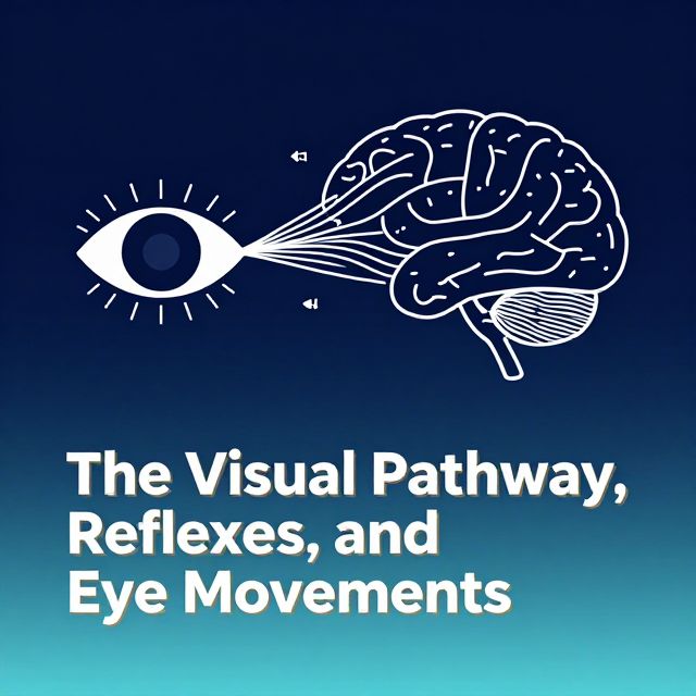
👁️ The Visual System: Pathway, Reflexes, and Eye Movements

1. Introduction to the Visual Pathway 📚

The visual system is a sophisticated network responsible for processing visual information, from initial light detection to complex interpretation in the brain. This guide details the anatomical route of visual signals, specialized processing streams, and reflex mechanisms controlling pupillary responses and eye movements.

2. The Visual Pathway: From Eye to Cortex 🧠

The journey of visual information begins in the retina and culminates in the primary visual cortex (V1).

2.1. Anatomical Route ✅

  1. Retina: Detects the visual field as an overturned (upside down) and right-left reversed image.
  2. Optic Nerve: Transmits signals from each eye.
  3. Optic Chiasm: Where fibers from the nasal (medial) half of each retina cross to the contralateral side.
  4. Optic Tract: Contains fibers from both eyes, carrying information from the contralateral visual field.
  5. Lateral Geniculate Body (LGB):
    • 90% of optic tract fibers project here. The LGB is a crucial relay station in the thalamus.
    • 10% of fibers bypass the LGB and project to other areas for specific functions:
      • Suprachiasmatic Nucleus: Involved in circadian rhythms.
      • Pretectal Nucleus: Essential for the light reflex.
      • Superior Colliculus: Controls eye movements and visual orientation.
      • Pineal Gland: Influences melatonin production.
  6. Optic Radiation (Tractus Geniculocalcarinus): Fibers extending from the LGB to the visual cortex.
    • Baum's Loop: Superior fibers, located in the parietal lobe.
    • Meyer's Loop: Inferior fibers, located in the temporal lobe.
  7. Primary Visual Cortex (V1): Also known as the striate cortex or Broadmann Area 17, located around the calcarine fissure in the occipital lobe.
    • Cuneus: Superior to the calcarine fissure.
    • Lingual Gyrus: Inferior to the calcarine fissure.

2.2. Visual Field and Cortical Representation 📊

The visual field is divided into zones:

  • Macular Zone: Responsible for central, high-acuity vision. It projects to the pole of the occipital cortex and occupies a proportionally larger surface area of the cortex compared to other zones.
    • 💡 Clinical Significance: The pole of the occipital cortex has a unique dual blood supply from both the Middle Cerebral Artery (MCA) and Posterior Cerebral Artery (PCA). This often leads to macular sparing in cases of PCA occlusion, meaning central vision may be preserved despite significant peripheral visual field loss.
  • Binocular Zone: Area seen by both eyes.
  • Monocular Zone: Area seen by only one eye (e.g., the far periphery).

Visual field deficits are termed:

  • Anopsias: Relatively large visual field deficits.
  • Scotomas: Smaller visual field deficits.
  • ⚠️ Possible Causes: Optic neuritis, central retinal artery occlusion, ICA aneurysm, pituitary adenoma, MCA/PCA occlusion.

3. Lateral Geniculate Body (LGB) and Visual Streams 📈

The LGB is a six-layered structure that segregates visual information.

3.1. LGB Lamination ✅

  • Layers 1, 4, 6: Receive fibers from the contralateral nasal retina.
  • Layers 2, 3, 5: Receive fibers from the ipsilateral temporal retina.
    • This segregation helps maintain the retinotopic map and prepares information for cortical processing.

3.2. Magno-, Parvo-, and Koniocellular Streams 🚀

These distinct pathways process different aspects of visual information.

| Stream | Layers Involved | % Fibers | Axon Type | Speed | Primary Function | Projection Target | | :--------------- | :-------------- | :------- | :-------- | :---- | :--------------------- | :--------------------- | | Magnocellular | 1, 2 | 10% | Thick | Fast | Movement, depth, flicker | Superior Colliculus, V1-4Cα | | Parvocellular | 3, 4, 5, 6 | 80% | Thin | Slow | Fine details, form, color | V1-4Cβ | | Koniocellular | Between layers | <10% | Very thin | Slow | Short-wavelength (S) cones (blue/yellow color) | V1-Layer 2 & 3 (patchy) |

4. Visual Cortex: Beyond V1 🎨

The visual cortex is organized into hierarchical areas for increasingly complex processing.

4.1. Primary Visual Cortex (V1) ✅

  • Also known as the striate cortex (Broadmann Area 17).
  • Layer 4: Receives axons directly from the LGB. Inputs from the two eyes remain segregated here before converging onto individual neurons in other cortical layers.
  • Line of Gennari: A visible band of myelinated axons from the LGB terminating in layer 4, seen in gross transverse sections.

4.2. Extrastriate Cortex (V2-V6) 🖼️

These areas (Broadmann Areas 18-19) process specific visual attributes:

  • V2, V3: Further processing of form and motion.
  • V4:
    • Function: Responds selectively to color and is responsible for high-resolution form vision and object recognition. Often considered part of the ventral stream ("what" pathway).
    • Lesion: Can cause cerebral achromatopsia (inability to perceive colors).
  • V5 (Middle Temporal Area - MT):
    • Function: Responds selectively to the direction of moving edges and is responsible for spatial aspects of vision, such as motion analysis and positional relationships. Often considered part of the dorsal stream ("where" pathway).
    • Lesion: Can cause cerebral akinetopsia (inability to perceive motion).
  • V6: Involved in motion and spatial orientation.

5. Visual Reflexes: Pupillary Control 💡

5.1. Light Reflex (Pupillary Light Reflex) 🔦

An involuntary constriction of the pupil in response to light.

  1. Light to Retina: Photoreceptors detect light.
  2. Optic Nerve: Transmits signals.
  3. Optic Tract: Signals travel bilaterally.
  4. Pretectal Nucleus: Receives input from both optic tracts.
  5. Edinger-Westphal (EW) Nucleus: The pretectal nucleus innervates both EW nuclei (parasympathetic).
  6. Ciliary Ganglion: EW nucleus fibers synapse here.
  7. Sphincter Pupillae Muscle: Postganglionic fibers cause contraction (miosis).
    • Direct Light Reflex: Constriction of the pupil in the eye stimulated by light.
    • Indirect (Consensual) Light Reflex: Constriction of the pupil in the unstimulated eye.
  • ⚠️ Important Note: Lesions in the optic radiation do not damage the light reflex because the pathway diverges at the optic tract, before the LGB and optic radiation.

5.2. Accommodation Reflex (Near Response) 🎯

Activated when eyes focus on close objects, involving three coordinated actions:

  1. Accommodation: Lens bulges (ciliary muscle contracts), controlled by the ventral part of the EW nucleus.
  2. Miosis: Pupil constricts (sphincter pupillae muscle contracts), controlled by the dorsal part of the EW nucleus.
  3. Convergence: Medial recti muscles contract, innervated by the oculomotor nucleus, which is stimulated by a convergence center near it.

5.3. Pupillary Abnormalities 🩺

  • Argyll-Robertson Pupil:
    • Cause: Often seen in neurosyphilis, affecting the dorsal part of the Edinger-Westphal nucleus.
    • Presentation: Absence of light reflex (pupil does not constrict to light) but preservation of accommodation reflex (pupil constricts during near vision). "Prostitute's pupil: accommodates but does not react."
  • Adie's Pupil:
    • Cause: Partial lesion of the ciliary ganglion.
    • Presentation: Loss of accommodation reflex (difficulty focusing on near objects) but a partially intact light reflex (slow, tonic constriction to light).
  • Marcus-Gunn Pupil (Relative Afferent Pupillary Defect - RAPD):
    • Cause: Unilateral or asymmetric optic nerve damage.
    • Presentation: When light is swung from the unaffected eye to the affected eye, the affected pupil paradoxically dilates or constricts less than expected, indicating a reduced afferent signal from that eye.

6. Conjugate Eye Movements: Gaze Control ↔️

The eyes normally move as a pair (conjugate movement).

6.1. Types of Eye Movements ✅

  • Gaze Shifting:
    • Saccadic Movements: Rapid, voluntary "jerk" movements to shift gaze to a new target.
    • Convergence: Involuntary movement of both eyes inward to focus on a near object.
  • Gaze Holding:
    • Tracking (Smooth Pursuit): Voluntary slow movements to follow a moving object.
    • Vestibulo-Ocular Reflex (VOR): Involuntary reflex that stabilizes gaze on an object during head movements.

6.2. Control Centers 🧠

  • Saccadic Movements (Searching Movements):
    • Cortical Center: Frontal Eye Field (FEF).
    • Horizontal Gaze: FEF controls the contralateral Paramedian Pontine Reticular Formation (PPRF). The PPRF controls the ipsilateral abducens nucleus (for lateral rectus) and the contralateral oculomotor nucleus (for medial rectus via the Medial Longitudinal Fasciculus - MLF).
    • Vertical Gaze: FEF controls nuclei in the dorsal mesencephalon (e.g., Rostral Interstitial Nucleus of MLF, Cajal Nucleus) bilaterally.
  • Tracking Movements (Smooth Pursuit):
    • Cortical Center: Occipital Eye Field (OEF).
    • The brainstem pathways are similar to saccadic movements but initiated by a different cortical area.

Kendi çalışma materyalini oluştur

PDF, YouTube videosu veya herhangi bir konuyu dakikalar içinde podcast, özet, flash kart ve quiz'e dönüştür. 1.000.000+ kullanıcı tercih ediyor.

Sıradaki Konular

Tümünü keşfet
Neuron Types and Nervous System Structures

Neuron Types and Nervous System Structures

This summary explores the diverse morphology and classification of neurons, their fundamental components, synaptic connections, and the crucial role of glial cells and peripheral nervous system structures.

6 dk Özet 25 15
Nervous System: Structure, Function, and Signaling

Nervous System: Structure, Function, and Signaling

An academic overview of the nervous system's anatomical and functional divisions, cellular components, electrical signaling mechanisms, and inter-neuronal communication.

5 dk Özet 25 15
Skeletal Muscle Structure and Neuromuscular Junction

Skeletal Muscle Structure and Neuromuscular Junction

This audio summary provides an academic overview of skeletal muscle terminology, morphology, filament and sarcomere structure, innervation, and the intricate process of excitation-contraction coupling.

7 dk Özet 18 12
Introduction to Visual Pathways and Lesions

Introduction to Visual Pathways and Lesions

Explore the visual pathway from the retina to the primary visual cortex, understand visual defects from lesions, and learn about post-V1 processing streams.

8 dk Özet 15
The Anatomy and Physiology of the Human Heart

The Anatomy and Physiology of the Human Heart

Explore the intricate structure and function of the human heart, covering its location, chambers, valves, and the electrical conduction system that drives its rhythmic beat.

Özet 25 15
The Vestibular System and Auditory Pathway

The Vestibular System and Auditory Pathway

An academic overview of the vestibular system's role in balance and spatial orientation, and the auditory pathway's function in sound perception, detailing their anatomical structures and neural processing.

8 dk Özet 25 15
Gastrointestinal Tract Blood Flow and Movements

Gastrointestinal Tract Blood Flow and Movements

Explore the intricate details of gastrointestinal blood circulation, including splanchnic flow, anatomical supply, regulatory factors, and the mechanics of propulsive and mixing movements within the GI tract.

9 dk Özet 15
Human Brain Anatomy: Ventricles, Terminology, and Structures

Human Brain Anatomy: Ventricles, Terminology, and Structures

Explore the intricate anatomy of the human brain, covering the ventricular system, essential directional terms, embryonic development, and the major divisions and structures of the adult brain.

25 15