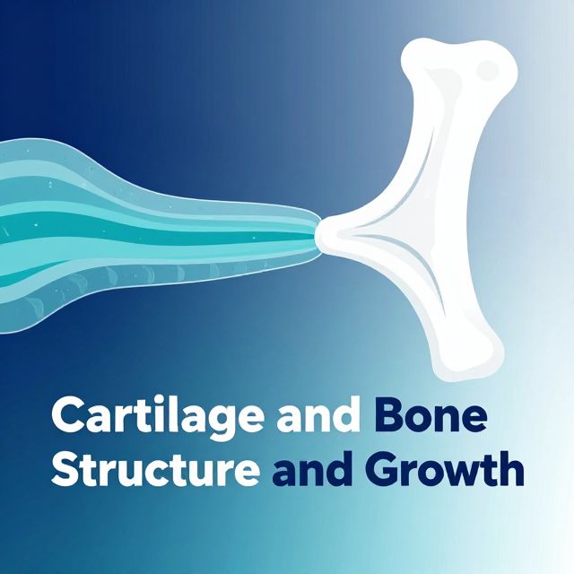
Comprehensive Study Guide: Cartilage and Bone Dynamics
This study material has been compiled and organized from various sources, including a lecture audio transcript and copy-pasted text, to provide a structured overview of cartilage and bone biology. It is designed for university-level students, particularly those preparing for medical faculty examinations, focusing on detailed explanations and key distinctions.
1. Introduction to Cartilage and Bone Dynamics 📚
Cartilage and bone are specialized connective tissues crucial for skeletal support, movement, and protection. This guide will explore their intricate structures, the composition of their extracellular matrices, their formation processes (chondrogenesis and ossification), and the distinct characteristics of different cartilage types. Understanding these fundamental biological processes is essential for comprehending skeletal development, maintenance, and pathology.
2. Cartilage Structure and Perichondrium 🦴
Cartilage is a resilient, semi-rigid form of connective tissue. Most cartilages are enveloped by a specialized dense connective tissue called the perichondrium.
2.1. Perichondrium Layers ✅
The perichondrium is an irregular dense connective tissue composed of two distinct layers:
- Outer Fibrous Layer:
- Primarily provides structural support and protection.
- Rich in Type I collagen fibers.
- Contains elastin fibers, contributing to flexibility.
- Houses fibroblasts, which are responsible for synthesizing the fibrous components.
- Inner Chondrogenic Layer:
- Located closer to the cartilage tissue.
- Contains chondrogenic cells, which are mesenchymal stem cells capable of differentiating into cartilage-forming cells.
- Crucial for cartilage growth and repair.
3. Extracellular Matrix (ECM) of Cartilage 💧
The ECM is the non-cellular component of cartilage, providing its unique mechanical properties. It is a complex mixture of fibers and ground substance.
3.1. Key Components of ECM ✅
- Hyaluronic acid: A large glycosaminoglycan (GAG) that provides hydration and acts as a backbone for proteoglycan aggregates.
- Glycosaminoglycans (GAGs): Long, unbranched polysaccharides that are highly negatively charged, attracting water.
- Proteoglycans: Core proteins to which GAGs are covalently attached. They form large aggregates (like aggrecan) that trap water.
- Aggrecan: The most abundant proteoglycan in cartilage, forming large aggregates with hyaluronic acid, which is critical for cartilage's ability to resist compression.
- Water (70-80%): The high water content, largely bound by proteoglycans, gives cartilage its turgor and resilience, allowing it to withstand compressive forces.
- Type II collagen: The predominant collagen type in most cartilages, forming a meshwork that provides tensile strength.
3.2. Organization of ECM 📊
The ECM is not uniformly distributed but organized into distinct regions around the chondrocytes:
- Pericellular Matrix: The immediate matrix surrounding each chondrocyte. It is rich in proteoglycans and glycoproteins, acting as a protective capsule.
- Territorial Matrix: Surrounds groups of chondrocytes (isogenous groups).
- Displays metachromasia: Stains a different color than the dye itself due to high concentrations of negatively charged GAGs.
- Stains positively with the PAS (Periodic Acid-Schiff) method, indicating the presence of carbohydrates.
- Interterritorial Matrix: The largest region, located between the territorial matrices and cell groups. It is less rich in proteoglycans than the territorial matrix.
4. Chondrogenesis: Cartilage Formation 📈
Cartilage develops through two primary mechanisms: interstitial and appositional growth.
4.1. Interstitial Growth 1️⃣
This type of growth occurs from within the cartilage mass.
- Step 1: Mesenchymal cells differentiate into chondrogenic cells.
- Step 2: Chondrogenic cells further differentiate into chondroblasts.
- Step 3: Chondroblasts actively synthesize and secrete the cartilage matrix.
- Step 4: As they become completely surrounded and entrapped by the matrix they produce, chondroblasts mature into chondrocytes.
- Step 5: Chondrocytes within the lacunae can divide, forming isogenous groups, and continue to produce matrix, expanding the cartilage from within. 💡 This growth is particularly important during early development and in the epiphyseal plates of long bones.
4.2. Appositional Growth 2️⃣
This growth occurs on the surface of pre-existing cartilage.
- Step 1: Chondrogenic cells in the inner layer of the perichondrium differentiate into chondroblasts.
- Step 2: These chondroblasts deposit new cartilage matrix onto the surface of the existing cartilage tissue.
- Step 3: As they become entrapped, they mature into chondrocytes. 💡 This process increases the width or thickness of the cartilage and is responsible for the growth of most cartilages throughout life.
5. Types of Cartilage and Their Characteristics 🔬
There are three main types of cartilage, each adapted to specific functions and locations.
5.1. Hyaline Cartilage 👃
- Characteristics:
- Most common type.
- ECM primarily contains Type II collagen.
- Smooth, glassy appearance.
- Locations:
- Forms the model for endochondral ossification in the fetus (precursor to bone).
- Found in the nose, trachea, bronchi, and most of the larynx.
- Covers the articular surfaces of synovial joints.
- Key Note: ⚠️ Joint cartilages (articular cartilage) do NOT have a perichondrium. This is crucial for their function in reducing friction and absorbing shock, as a perichondrium would hinder nutrient diffusion.
5.2. Elastic Cartilage 👂
- Characteristics:
- Highly flexible due to the presence of elastic fibers.
- Contains Type II collagen in its matrix.
- Possesses a perichondrium.
- Special Staining: Elastic fibers are visible with specific stains like Verhoeff, orcein, or resorcin-fuchsin.
- Locations:
- Auricle (external ear)
- Eustachian tube (auditory tube)
- Epiglottis
- Some laryngeal cartilages (e.g., cuneiform, corniculate, epiglottis).
5.3. Fibrous Cartilage (Fibrocartilage) 💪
- Characteristics:
- Strongest and most durable type of cartilage, designed to resist compression and shear forces.
- LACKS a perichondrium.
- Composed of both chondrocytes (often arranged in rows) and fibroblasts.
- ECM is rich in thick bundles of Type I collagen fibers.
- Contains fewer proteoglycans and less water compared to hyaline or elastic cartilage, contributing to its toughness.
- Locations:
- Intervertebral discs (annulus fibrosus)
- Symphysis pubis
- Temporomandibular joint (TMJ) disc
- Sternoclavicular joint disc
- Menisci of the knee joint.
6. Bone Growth Mechanisms 🦴
Bone growth involves both longitudinal lengthening and appositional thickening.
6.1. Epiphysial Plate (Growth Plate) 📏
The epiphysial plate is a hyaline cartilage structure responsible for the longitudinal growth of long bones.
- Function: Allows bones to lengthen during childhood and adolescence.
- Key Processes:
- Ossification occurs at the diaphysial side (side closer to the shaft of the bone).
- Mitosis of chondrocytes occurs at the epiphysial side (side closer to the end of the bone).
- Zones of the Epiphysial Plate (from epiphysis to diaphysis):
- Resting (Reserve) Zone: Chondrocytes are small, scattered, and relatively inactive. Serves as a reservoir of chondrocytes.
- Proliferation Zone: Chondrocytes undergo rapid mitosis, forming columns of cells. This pushes the epiphysis away from the diaphysis, lengthening the bone.
- Maturation (Hypertrophy) Zone: Chondrocytes enlarge significantly (hypertrophy), accumulating glycogen and secreting alkaline phosphatase. The lacunae also enlarge.
- Calcification Zone: The hypertrophied chondrocytes die, and the surrounding cartilage matrix becomes calcified by the deposition of calcium salts.
- Ossification Zone: Blood vessels invade the calcified cartilage. Osteoprogenitor cells differentiate into osteoblasts, which deposit new bone matrix on the calcified cartilage remnants, forming new bone tissue.
6.2. Thickening of Bone (Appositional Growth) 🏗️
Bone thickening, or an increase in diameter, occurs through appositional growth.
- Mechanism:
- Osteoprogenitor cells located in the inner layer of the periosteum (the connective tissue covering the outer surface of bone) differentiate into osteoblasts.
- These osteoblasts synthesize and secrete new bone matrix (osteoid) onto the existing bone surface.
- As they become entrapped within this newly formed matrix, they mature into osteocytes.
- This process adds new bone tissue to the outer surface, specifically in the subperiosteal zone, increasing the bone's diameter.
- Concurrent Process: While new bone is added externally, the inner aspect of the bone tissue is eroded by osteoclasts. This process enlarges the bone marrow cavity, maintaining a balance between bone formation and resorption, and optimizing bone structure for strength without excessive weight.
Conclusion ✅
This study guide has provided a detailed examination of cartilage and bone, covering their structural components, ECM organization, and growth mechanisms. Understanding the perichondrium, the distinct types of cartilage (hyaline, elastic, fibrocartilage) with their specific collagen types and perichondrial presence, and the processes of chondrogenesis and bone growth (epiphysial plate and appositional thickening) is fundamental for a comprehensive grasp of musculoskeletal biology. These intricate processes collectively ensure the development, maintenance, and adaptive capacity of the skeletal system, which is vital for health and function.