Understanding Animal Tissues: Structure, Function, and Development - kapak
Bilim#animal tissues#epithelium#connective tissue#histology

Understanding Animal Tissues: Structure, Function, and Development

Explore the intricate world of animal tissues, focusing on epithelium and connective tissues. Learn about their embryonic origins, diverse structures, and vital functions in the body.

banoJanuary 22, 2026 ~25 dk toplam
01

Sesli Özet

9 dakika

Konuyu otobüste, koşarken, yolda dinleyerek öğren.

Sesli Özet

Understanding Animal Tissues: Structure, Function, and Development

0:008:44
02

Flash Kartlar

25 kart

Karta tıklayarak çevir. ← → ile gez, ⎵ ile çevir.

1 / 25
Tüm kartları metin olarak gör
  1. 1. What is the definition of a tissue in biology?

    Tissues are defined as groups of cells that differentiate similarly to perform a specific function. This organization allows multicellular organisms to carry out complex biological processes efficiently. The study of these tissues is known as histology.

  2. 2. What is histology?

    Histology is the scientific field dedicated to the examination and study of tissues. It involves observing the microscopic structure of tissues to understand their organization, function, and how they contribute to the overall physiology of an organism.

  3. 3. Name the three primary embryonic layers from which all animal tissues originate.

    All animal tissues originate from three primary embryonic layers: the ectoderm, mesoderm, and endoderm. These layers form during early embryonic development and give rise to distinct sets of tissues and organs, determining their specialized roles.

  4. 4. Which embryonic layer primarily gives rise to nerve tissue and epithelium covering the external body surface?

    Nerve tissue and the epithelium covering the external body surface primarily originate from the ectoderm. The ectoderm is the outermost germ layer, responsible for forming structures that interact with the external environment, including the nervous system and skin.

  5. 5. From which embryonic layer do connective tissue, muscle tissue, and blood tissue primarily originate?

    Connective tissue, muscle tissue, and blood tissue primarily originate from the mesoderm. The mesoderm is the middle germ layer, responsible for forming many of the body's structural and circulatory components, including muscles, bones, and blood.

  6. 6. What are the general characteristics of epithelium tissue regarding cell arrangement and vascularity?

    Epithelium tissue is characterized by cells arranged in a regular, side-by-side manner, forming continuous sheets. A key characteristic is its avascular nature, meaning it lacks blood vessels. Nutrients are supplied by diffusion from underlying connective tissue.

  7. 7. How does the structure of epithelium tissue differ between invertebrates and vertebrates?

    Structurally, epithelium tissue is typically single-layered in invertebrates. In contrast, vertebrates possess multi-layered epithelium. This difference reflects the increased complexity and protective needs of vertebrate organisms.

  8. 8. List four general functions of epithelium tissue.

    Four general functions of epithelium tissue include protection (e.g., skin), secretion (e.g., by glands), acting as receptors (e.g., in sense organs), and absorption (e.g., in the intestines). These diverse roles highlight its importance in various physiological processes.

  9. 9. What are the three main types of epithelium tissue based on classification?

    Epithelium tissue can be categorized into three main types: covering and lining epithelium, glandular epithelium, and sensory epithelium. This classification helps to understand their distinct structures and specialized functions within the body.

  10. 10. Describe the function of covering and lining epithelium and provide an example.

    Covering and lining epithelium protects organs from external impacts and forms barriers. Examples include the epidermis of the skin, which protects the body surface, and the mucosa of the respiratory and digestive tracts, which line internal passages.

  11. 11. How do glandular epithelium cells form and what is the difference between 'secretion' (substance) and 'secretion' (process)?

    Glandular epithelium forms from altered cubic or cylindrical cells. The substance accumulated within these cells is called 'secretion,' while its release from the cell is also termed 'secretion.' These substances often include enzymes or mucous.

  12. 12. Explain the difference between exocrine and endocrine glands.

    Exocrine glands release their secretions through ducts onto an epithelial surface or into a cavity, such as sweat glands or salivary glands. Endocrine glands, however, lack ducts and release their secretions (hormones) directly into the bloodstream for systemic distribution.

  13. 13. Provide an example for each of the three types of exocrine secretion: merocrine, apocrine, and holocrine.

    Merocrine secretion involves the cell remaining intact after releasing its contents, like in sweat glands. Apocrine secretion releases a portion of the cytoplasm with the secretion, as seen in armpit glands. Holocrine secretion involves the entire cell becoming the secretion, exemplified by lipocytes.

  14. 14. What are mixed glands, and can you give an example?

    Mixed glands are organs that possess both exocrine and endocrine functions, producing both enzymes and hormones. A prime example is the pancreas, which produces digestive enzymes (exocrine) and hormones like insulin and glucagon (endocrine). The stomach also functions as a mixed gland.

  15. 15. What is the primary function of sensory epithelium?

    Sensory epithelium forms the receptors of our five sense organs. Its primary function is to detect specific stimuli from the environment, such as light, sound, taste, smell, and touch, and convert them into nerve signals for interpretation by the nervous system.

  16. 16. What is the embryonic origin of connective tissue, and what is its prevalence in the body?

    Connective tissue originates from the mesoderm layer of the embryo. It is the most common tissue found throughout the organism, playing crucial roles in support, binding, and protection for various body structures.

  17. 17. What is the distinguishing feature of connective tissue compared to epithelial tissues, regarding cell count and interstitial substance?

    Connective tissue is distinguished from epithelial tissues by its lower cell count and a higher amount of interstitial substance, known as the 'matrix.' This matrix is critically important and varies significantly in structure depending on the type of connective tissue.

  18. 18. Name the three main types of connective tissue discussed.

    The three main types of connective tissue discussed are loose connective tissue, cartilage tissue, and bone tissue. Each type has specialized structures and functions, contributing to the body's overall support and integrity.

  19. 19. What are the primary cells in loose connective tissue, and what are its main components?

    The primary cells in loose connective tissue are fibroblasts. Its matrix consists of proteins, polysaccharides, and minerals. Loose connective tissue loosely binds structures, surrounds blood vessels and nerves, and connects muscles to muscles and bones to bones.

  20. 20. Describe adipose tissue, its specialized cells, and its different types.

    Adipose tissue is a specialized form of loose connective tissue comprising adipocytes that store triglycerides. It can appear brown, white, or rarely yellow. Brown adipose tissue is more prevalent in embryos and newborns, while adults primarily have white adipose tissue.

  21. 21. List three vital functions of adipose tissue.

    Three vital functions of adipose tissue include serving as an energy source, acting as an insulator against heat loss, and filling spaces between organs to help them maintain shape. It also functions as a buffer against impacts during falls.

  22. 22. What is the role of cartilage tissue during the embryonic stage of vertebrates, and where does it primarily remain in adulthood?

    Cartilage tissue forms the entire skeleton during the embryonic stage of all vertebrates. In adulthood, as bone tissue increases, cartilage remains primarily in joints, ribs, ear lobes, and the nose, providing flexibility and support.

  23. 23. What are three key functions of cartilage tissue?

    Three key functions of cartilage tissue include serving as the embryonic skeleton, enabling free movement of joints, and supporting soft tissues such as the ear, nose, trachea, and bronchus. It also plays a role in transforming into bone tissue.

  24. 24. What is the hardest tissue in our bodies, and what is its exclusivity regarding animal groups?

    Bone tissue is the hardest tissue in our bodies, forming the skeleton. It is exclusive to vertebrates, developing from cartilage tissue in embryos, and provides structural support, protection, and facilitates movement.

  25. 25. What are the main components of the bone tissue matrix, and what functions do organic and inorganic matter provide?

    The bone tissue matrix is composed of water, organic matter (ossein and collagen), and inorganic matter (calcium salts like CaCO3 and Ca3(PO4)2). Organic matter provides elasticity, preventing easy fractures, while inorganic matter gives bones their hardness.

03

Bilgini Test Et

15 soru

Çoktan seçmeli sorularla öğrendiklerini ölç. Cevap + açıklama.

Soru 1 / 15Skor: 0

What is the scientific field dedicated to examining tissues?

04

Detaylı Özet

6 dk okuma

Tüm konuyu derinlemesine, başlık başlık.

📚 Animal Tissues: Structure, Function, and Embryonic Origins 📚

Source Information: This study material has been compiled from a copy-pasted text provided by the user and a lecture audio transcript.


🎯 Introduction to Animal Tissues

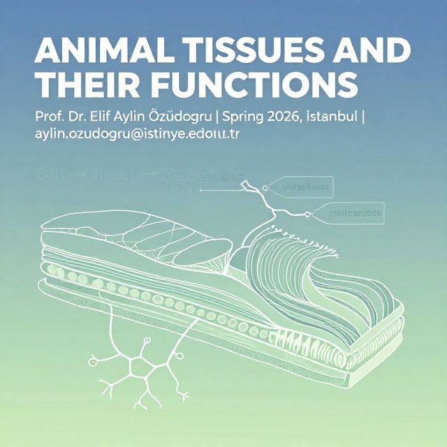
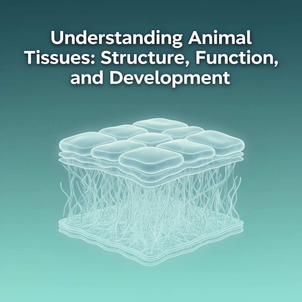
Animal tissues are fundamental building blocks of multicellular organisms, comprising groups of cells that differentiate similarly to perform specific functions. The scientific study of tissues is known as histology. This guide focuses exclusively on animal tissues, exploring their diverse structures, specialized functions, and embryonic origins.


🧬 Embryonic Origins of Tissues

All animal tissues originate from the three primary germ layers formed during embryonic development: the ectoderm, mesoderm, and endoderm. During the embryonic stage, cell groups from these layers differentiate to form all the various tissues of the body.

Five Basic Tissue Types and Their Primary Origins:

  1. Epithelium Tissue: Primarily originates from the ectoderm, but also from mesoderm and endoderm.
  2. Connective Tissue: Originates from the mesoderm.
  3. Muscle Tissue: Originates from the mesoderm.
  4. Blood Tissue: Originates from the mesoderm.
  5. Nerve Tissue: Originates from the ectoderm.

1️⃣ Epithelium Tissue

Epithelium tissue forms coverings and linings throughout the body, including external surfaces, internal organs, blood vessels, and all body cavities.

🔬 Characteristics and Structure:

  • Cell Arrangement: Cells are regularly arranged, side-by-side.
  • Intercellular Spaces: Small spaces (approx. 80Aº) exist between cells, filled with interstitial fluid. Cell membranes do not directly touch.
  • Vascularity: Lacks blood vessels.
  • Layering: Single-layered in invertebrates; multi-layered in vertebrates.

embryological Development:

Epithelium tissue is versatile in its embryonic origin:

  • Ectoderm: Forms epithelium covering the external surface of the body (e.g., skin epidermis).
  • Mesoderm: Forms epithelium covering kidneys and genitalia.
  • Endoderm: Forms epithelium lining the internal surface of the digestive tract.

🛡️ General Functions:

Epithelium tissue is flexible and provides crucial protection. Its main functions include:

  • Protection: Against chemical, mechanical, and physical effects (e.g., skin).
  • Secretion: Production and release of substances (e.g., secretory glands).
  • Reception: Sensory input (e.g., sense organs).
  • Absorption: Uptake of substances (e.g., intestines).

📊 Classification of Epithelium Tissue:

Epithelium is named and grouped based on cell layering, shapes, and functions.

a) Covering and Lining Epithelium:

This type covers external organ surfaces and protects them from external impacts.

  • Examples:
    • Epidermis: Outermost layer of the skin.
    • Conjunctiva Epithelium: Internal surface of the eyelids.
    • Mucosa Epithelium: Internal surfaces of the respiratory and digestive tracts.
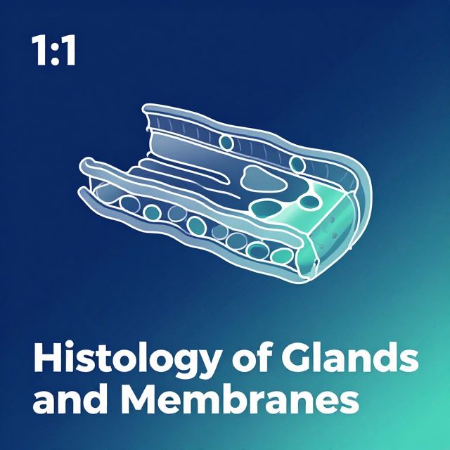
b) Glandular Epithelium:

Formed by modified cubic or cylindrical epithelial cells, specialized for secretion.

  • Secretion: The substance accumulated within the cell.
  • Secretion Process: The release of this substance outside the cell.
  • Secretions: Can be enzymes (e.g., for digestion) or mucous (e.g., for moisture and lubrication).
    • 💡 Example: Mucosa cells in frogs and worms moisten skin for cutaneous respiration.
3️⃣ Types of Secretory Glands (by secretion release location):
  1. Exocrine Glands: Release secretions into ducts that lead to an external or internal surface. Can be unicellular or multicellular.
    • Merocrine Secretion: Secretion is released from a slit on the cell surface; the cell remains largely intact.
      • Examples: Sweat glands, mucous glands of digestive/respiratory systems, pancreas, kidney.
    • Apocrine Secretion: A portion of the cytoplasm is released along with the secretion; the nucleus remains intact for cell repair.
      • Examples: Glands in armpit, earwax, inguinal region, scrotum, around anus.
    • Holocrine Secretion: The entire cell disintegrates and becomes the secretion; reserve cells replace lost cells.
      • Example: Lipocytes (though often associated with sebaceous glands).
  2. Endocrine Glands: Lack secretory channels; release secretions (called incretes or hormones) directly into the blood circulation. This process is called incretion.
    • Examples: Pituitary gland, epiphysis, thyroid, parathyroid, adrenal gland, pancreas (endocrine part), sexual glands. Hormones are typically released via exocytosis.
  3. Mixed Glands: Possess both exocrine and endocrine functions.
    • Examples: Pancreas (produces digestive enzymes like amylase, lipase, and hormones like insulin, glucagon), stomach.
c) Sensory Epithelium:

Specialized epithelial cells that act as receptors for the five sense organs.

✅ Comprehensive Functions of Epithelium Tissue:

  • i) Protection: Shields the organism from external impacts and microorganisms.
  • ii) Absorption: Facilitates nutrient uptake (e.g., in intestines).
  • iii) Secretion: Produces and releases substances (e.g., sweat, saliva).
  • iv) Sense: Contains receptors for sensory perception.
  • v) Contraction: Some specialized epithelial cells provide mechanical aid.
  • vi) Extraction: Involved in waste discharge (e.g., from kidneys).
  • vii) Transport: Covers capillary vessels, enabling substance exchange between blood and cells.

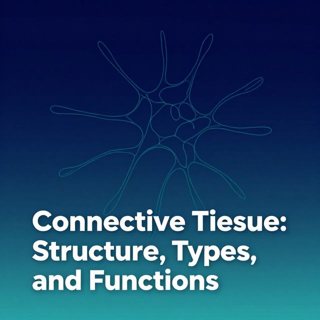
2️⃣ Connective Tissue

Connective tissue is the most common tissue type in the body, originating from the mesoderm. It provides support, binds structures, and fills spaces.

🔬 Characteristics:

  • Vascularity: Contains blood vessels and nerves.
  • Location: Surrounds epithelial cells, spaces between muscle cells, and blood vessels.
  • Composition: Characterized by fewer cells and a larger amount of interstitial substance (or matrix).
  • Matrix: Crucial for tissue function; its structure varies significantly by connective tissue type.
  • Supportive Role: Also known as supportive tissue, alongside bone and cartilage.

📊 Types of Connective Tissue:

a) Loose Connective Tissue:

Loosely binds body structures, keeping tissues in their normal locations.

  • Location: Surrounds blood vessels and nerves, connects muscles to muscles, and bones to bones.
  • Cells: Primarily fibroblasts.
  • Matrix: Fills spaces between fibroblasts, composed of proteins, polysaccharides, and minerals.
Adipose Tissue (Fat Tissue):

A specialized form of loose connective tissue.

  • Cells: Special connective tissue cells (adipocytes) that store fat.
  • Fat Storage: Adipocytes convert lipoproteins from the liver into triglycerides and store them.
  • Appearance: Can be Brown, White, or rarely Yellow.
    • Brown Adipose Tissue: Seen in embryos and newborns; generally absent in adults.
    • White Adipose Tissue: Predominant in adults.
  • Cell Development: Young cells are lipoblasts; mature, fat-filled cells are lipocytes (adipocytes).
  • Functions:
    • Energy Source: Stores energy.
    • Insulator: Provides thermal insulation.
    • Space Filler: Fills spaces between organs, helping them maintain shape.
    • Buffer: Acts as a shock absorber against impacts (e.g., during falls).
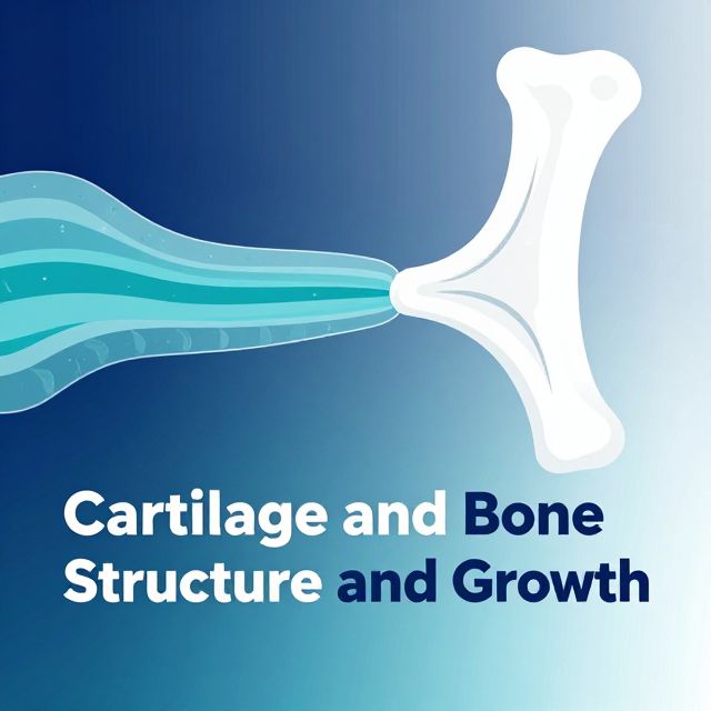
b) Cartilage Tissue:

A specialized connective tissue.

  • Embryonic Role: Forms the entire skeleton of vertebrates during the embryonic stage.
  • Adult Distribution: Decreases in adulthood as bone tissue increases; remains in joints, ribs, ear lobe, and nose.
    • 💡 Example: Primitive vertebrates like sharks retain a cartilaginous skeleton throughout life.
  • Functions:
    • Skeletal Support: Serves as the embryonic skeleton.
    • Joint Movement: Enables free movement of joints, providing bone articulation.
    • Soft Tissue Support: Supports structures like the ear, nose, trachea, and bronchi.
    • Bone Precursor: Can transform into bone tissue.
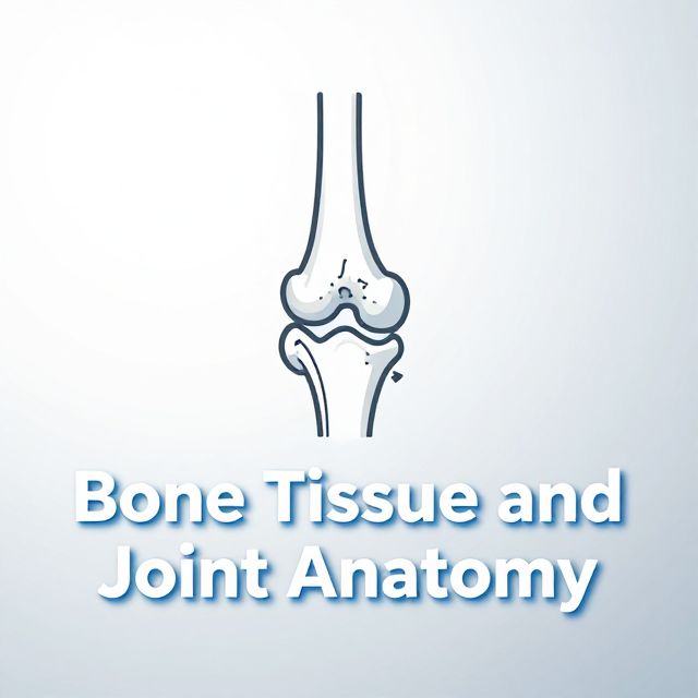
c) Bone Tissue:

The third type of connective tissue and the hardest tissue in the body, forming the skeleton.

  • Vertebrate Specific: Found only in vertebrates.
  • Development: Forms from cartilage tissue in embryos.
  • Muscle Connection: Generally bound to muscles by tendons, facilitating movement upon muscle contraction.
  • Matrix Composition (Adult):
    • Water: 17%
    • Organic Matter (27%): Ossein and collagen fibers, providing elasticity and preventing easy fractures.
    • Inorganic Matter (56%): Calcium salts (CaCO3, Ca3(PO4)2, calcium fluoride), providing hardness.
  • ⚠️ Aging Effects: With age, inorganic matter increases, and organic matter decreases, making bones harder, more brittle, prone to fractures, and slower to heal.
  • Rickets: Insufficiency of inorganic salts leads to bone softening and skeletal bending.
    • Treatment: Vitamins C (for bone structure) and D (for calcium absorption).
  • Bone Marrow: Soft tissue filling spaces within bones.
    • Red Marrow: Found in the tips of long bones; produces erythrocytes (red blood cells).
    • Yellow Marrow: Soft, stores fat; found in the middle of long bones in adults.
  • Functions:
    • Skeletal Formation: Forms the body's framework.
    • Movement: Facilitates movement in conjunction with muscles.
    • Organ Protection: Protects vital organs (e.g., brain, heart).
    • Calcium Source: Acts as a reservoir for calcium.
    • Hematopoiesis: Bone marrow produces red blood cells.

Kendi çalışma materyalini oluştur

PDF, YouTube videosu veya herhangi bir konuyu dakikalar içinde podcast, özet, flash kart ve quiz'e dönüştür. 1.000.000+ kullanıcı tercih ediyor.

Sıradaki Konular

Tümünü keşfet
Animal Tissues and Their Functions in Vertebrates

Animal Tissues and Their Functions in Vertebrates

An educational overview of the four primary animal tissues in vertebrates: epithelial, connective, muscle, and nervous tissues, including their structure, functions, and locations.

Özet 25 15
Understanding Connective Tissues and Their Cells

Understanding Connective Tissues and Their Cells

Explore the fundamental types, structures, and cellular components of connective tissues, including their crucial roles in the body's support, protection, and immunity.

Özet 25 15
Connective Tissue Cells: Structure, Function, and Roles

Connective Tissue Cells: Structure, Function, and Roles

This summary provides an academic overview of connective tissue cells, detailing their classification, morphology, specific functions, and critical roles in tissue maintenance, repair, and immune responses.

9 dk Özet 25 15
Connective Tissue: Structure, Types, and Functions

Connective Tissue: Structure, Types, and Functions

An academic overview of connective tissue, detailing its embryonic origins, classifications into proper and specialized types, and the composition and roles of its fibrous and extracellular matrix components.

7 dk Özet 25 15
Histology of Glands and Membranes

Histology of Glands and Membranes

An academic overview of glandular classification, secretion mechanisms, and the structural and functional characteristics of various biological membranes in histology.

5 dk Özet 25 15
Cartilage and Bone Structure and Growth

Cartilage and Bone Structure and Growth

This summary provides an academic overview of cartilage structure, extracellular matrix, chondrogenesis, distinct cartilage types, and the mechanisms of bone growth and thickening.

6 dk Özet 25 15
Bone Tissue and Joint Anatomy

Bone Tissue and Joint Anatomy

An academic summary detailing the composition, structure, cellular components, development, and pathologies of bone tissue, alongside an overview of joint types and synovial joint anatomy.

14 dk Özet 25 15
Understanding the Borehole Environment

Understanding the Borehole Environment

This summary provides an academic overview of the borehole environment, detailing its characteristics, influencing factors, and significance in subsurface investigations and resource extraction.

6 dk Özet 15