Animal Tissues and Their Functions in Vertebrates - kapak
Bilim#animal tissues#vertebrate anatomy#epithelial tissue#connective tissue

Animal Tissues and Their Functions in Vertebrates

An educational overview of the four primary animal tissues in vertebrates: epithelial, connective, muscle, and nervous tissues, including their structure, functions, and locations.

izemFebruary 21, 2026 ~17 dk toplam
01

Flash Kartlar

25 kart

Karta tıklayarak çevir. ← → ile gez, ⎵ ile çevir.

1 / 25
Tüm kartları metin olarak gör
  1. 1. Describe the four levels of organizational hierarchy in vertebrate bodies.

    Vertebrate bodies exhibit a sophisticated organizational hierarchy starting with individual cells. These cells then aggregate to form tissues, which are groups of similar cells with shared functions. Tissues combine to create organs, which are structural and functional units made of several different tissues. Finally, multiple organs collaborate within organ systems to execute major activities essential for survival.

  2. 2. What is the definition of an organ in the context of vertebrate organization?

    An organ is defined as a body structure composed of several different tissues that collectively form a structural and functional unit. For example, the stomach is an organ made of epithelial, connective, muscle, and nervous tissues working together to digest food. Its distinct structure allows it to perform a specific physiological role.

  3. 3. Name the three fundamental embryonic tissues, also known as germ layers, from which all other tissues differentiate.

    The three fundamental embryonic tissues, or germ layers, are the endoderm, mesoderm, and ectoderm. These layers are formed early in embryonic development. They are crucial because all distinct cell types and primary tissues characteristic of the vertebrate body differentiate from these initial germ layers.

  4. 4. What are the four primary tissue types found in the vertebrate body?

    The four primary tissue types found in the vertebrate body are epithelial, connective, muscle, and nervous tissues. Each of these tissue types has specialized structures and functions. They work together to form organs and organ systems, enabling the complex physiological processes of vertebrates.

  5. 5. Where can epithelial tissue be found in the body, and what are its main functions?

    Epithelial tissue is found in various crucial locations, including body coverings, body linings, and glandular tissue. Its main functions are diverse, encompassing barrier formation, protection against external factors, absorption of substances, filtration of fluids, and secretion of various compounds. It essentially covers virtually every surface of the vertebrate body.

  6. 6. Explain a key characteristic of epithelial tissue regarding its cell arrangement and intercellular space.

    A key characteristic of epithelial tissue is that its cells are tightly bound together, with very little intercellular space between them. This tight packing allows epithelial tissue to form effective barriers and linings. This arrangement also contributes to its functions in protection and regulation of substance passage.

  7. 7. How do epithelial cells receive nutrients and oxygen, and what is a consequence of this method?

    Epithelial cells receive nutrients and oxygen via diffusion from blood vessels located in adjacent tissues. This method of nutrient supply means that epithelial tissue lacks its own direct blood supply. Consequently, this limits their thickness, typically to one or a few cell layers, as diffusion is only efficient over short distances.

  8. 8. Describe the regenerative potential of epithelial tissue, providing examples from the text.

    Epithelial tissue possesses remarkable regenerative potential, constantly replacing its cells throughout an organism's life. This high turnover rate is essential for tissues that experience wear and tear or are involved in secretion. Examples include the liver, which regenerates every two weeks, and the stomach lining, which renews every two to three days.

  9. 9. Differentiate between simple and stratified epithelial tissues based on their structure.

    Simple epithelial tissues are characterized by being one cell layer thick. An example is the flattened cells lining the lungs and blood capillaries, which permit rapid molecular movement. In contrast, stratified epithelial tissues are several cell layers thick and are named according to the features of their uppermost layers, providing greater protection.

  10. 10. What are the two main types of vertebrate glands, and how do they differ in their connection to the epithelial membrane and secretion method?

    The two main types of vertebrate glands are exocrine and endocrine glands. Exocrine glands maintain a connection to the epithelial membrane via a duct, secreting substances like sweat onto surfaces or digestive enzymes into tracts. Endocrine glands, however, are ductless; their secretions (hormones) enter blood capillaries directly, circulating within the body.

  11. 11. Provide examples of secretions from exocrine glands mentioned in the text.

    Exocrine glands secrete substances through ducts onto epithelial surfaces or into cavities. Examples mentioned in the text include sweat, which is secreted onto the skin's external surface. Additionally, digestive enzymes from salivary glands, the liver, and the pancreas are secreted into the digestive tract, aiding in food breakdown.

  12. 12. What is the primary characteristic that defines connective tissue, and how does its matrix vary?

    A defining feature of connective tissue is its abundant extracellular material, or matrix, which surrounds widely spaced cells. This matrix is crucial for its diverse functions and can vary greatly in consistency. Examples of this variation include the hard crystals found in bone and the fluid plasma that constitutes the matrix in blood.

  13. 13. List at least five functions of connective tissue in the body.

    Connective tissue performs numerous vital functions throughout the body. These include providing structural support and protection for organs, binding different structures together, filling spaces within the body, and storing fat for energy reserves. It also plays roles in producing blood cells, protecting against infection, and repairing tissue damage.

  14. 14. What are the two major classes of connective tissue?

    Connective tissues are broadly divided into two major classes: connective tissue proper and special connective tissues. Connective tissue proper includes types like loose and dense connective tissues, which are involved in binding and support. Special connective tissues, on the other hand, encompass more specialized forms such as cartilage, bone, and blood, each with unique properties.

  15. 15. Describe the characteristics and functions of loose connective tissue, mentioning its components.

    Loose connective tissue, such as areolar, reticular, and adipose tissue, is characterized by its loosely arranged fibers and cells. It functions to hold organs in place and attach epithelial tissue to underlying structures, providing flexibility and support. It contains cells that secrete collagen, mast cells producing histamine and heparin, and macrophages for immune defense.

  16. 16. What is adipose tissue composed of, and what is its primary function?

    Adipose tissue is a type of loose connective tissue primarily composed of adipose cells. These specialized cells are designed to store large fat droplets, specifically triglycerides. Its primary function is to serve as an energy reserve, as these stored triglycerides can be hydrolyzed and utilized for metabolic energy when needed by the body.

  17. 17. How does dense connective tissue differ from loose connective tissue in terms of structure and strength?

    Dense connective tissue is characterized by tightly packed collagen fibers, making it significantly stronger and more resistant to tension than loose connective tissue. In contrast, loose connective tissue has a more open, less fibrous structure. The high density of collagen in dense connective tissue provides robust structural support and tensile strength.

  18. 18. Differentiate between regular and irregular forms of dense connective tissue, providing an example for each.

    Dense connective tissue comes in regular and irregular forms. In regular dense connective tissue, collagen fibers are arranged parallel to each other, providing strength in one direction, as seen in tendons (binding muscle to bone) and ligaments (binding bone to bone). Irregular dense connective tissue has fibers with different orientations, providing strength in multiple directions, found in organ capsules like those of the kidneys.

  19. 19. Name the three types of special connective tissues mentioned in the text.

    The three types of special connective tissues mentioned in the text are cartilage, bone, and blood. These tissues are considered 'special' because they have unique compositions and highly specialized functions that distinguish them from connective tissue proper. Each plays a critical role in the body's structure, transport, and support.

  20. 20. What is the primary responsibility of muscle tissue, and what other vital roles does it play?

    The primary responsibility of muscle tissue is motion, facilitating both the movement of the entire body and localized movements of specific parts. Beyond movement, muscle tissue also plays vital roles in maintaining posture, helping to hold the body upright against gravity. Additionally, it contributes significantly to heat production during contraction, which helps regulate body temperature.

  21. 21. Which types of muscle are known as striated muscles, and why?

    Skeletal and cardiac muscles are known as striated muscles. They are called 'striated' because they exhibit characteristic transverse stripes or bands when viewed under a microscope. These striations are due to the regular arrangement of contractile proteins (actin and myosin) within their muscle fibers, which is essential for their contraction mechanism.

  22. 22. Differentiate between voluntary and involuntary control in muscle contraction, giving examples of muscle types for each.

    Voluntary control refers to muscle contractions that an individual can consciously direct, such as skeletal muscle contractions used for movement. In contrast, involuntary control refers to contractions that occur without conscious thought or effort. Cardiac muscle and smooth muscle contractions are generally involuntary, regulating functions like heartbeats and digestion automatically.

  23. 23. Describe the characteristics and location of smooth muscle tissue.

    Smooth muscle tissue is found in the internal organs, or viscera, such as the walls of the digestive tract, blood vessels, and bladder. Its cells are long, spindle-shaped, and contain a single nucleus. Smooth muscle contractions are generally involuntary, and in some tissues, they can spontaneously initiate electrical impulses, leading to slow, steady contractions.

  24. 24. How is skeletal muscle attached to bones, and what is its primary function?

    Skeletal muscle is attached to bones by strong, fibrous connective tissues called tendons. Its primary function is to produce all movements of body parts, from walking to lifting objects. It consists of numerous, very long muscle cells (muscle fibers) arranged parallel to each other, which are stimulated by nerve fibers to allow for varied contraction strength.

  25. 25. Describe the unique structural features of cardiac muscle and how they contribute to its function.

    Cardiac muscle, found exclusively in the heart, is striated but comprises smaller, interconnected cells, each with a single nucleus. These cells are linked by specialized structures called gap junctions, which allow electrical impulses to spread rapidly between them. This interconnection enables the cardiac muscle cells to form a single, functional unit (myocardium) and contract in a coordinated manner, essential for pumping blood.

02

Bilgini Test Et

15 soru

Çoktan seçmeli sorularla öğrendiklerini ölç. Cevap + açıklama.

Soru 1 / 15Skor: 0

What is the correct hierarchical organization of vertebrates, starting from the most basic functional unit?

03

Detaylı Özet

7 dk okuma

Tüm konuyu derinlemesine, başlık başlık.

Study Material: Animal Tissues and Their Functions

Source Information: This study material has been compiled from a lecture audio transcript and copy-pasted text, including presentation slides, provided by Prof. Dr. Elif Aylin Özüdoğru, Spring 2026, İstanbul.


📚 Introduction to Vertebrate Organization

The bodies of vertebrates exhibit a sophisticated organizational hierarchy, essential for complex physiological processes. This organization progresses through four distinct levels:

  1. Cells: The fundamental units of life.
  2. Tissues: Groups of similar cells working together.
  3. Organs: Structures composed of several different tissues.
  4. Organ Systems: Groups of organs functioning collaboratively.

Organs are body structures made up of multiple different tissues that form a structural and functional unit. ✅ An Organ System is a collection of organs that work together to perform major activities vital for the body's survival.

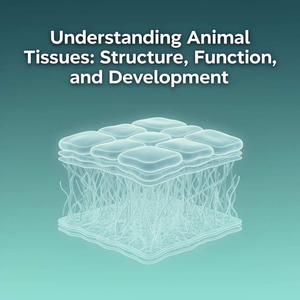
🧬 Tissues: The Building Blocks

At the foundational level, tissues are defined as groups of cells that share similar structure and function.

Early Embryonic Development: Early in development, the cells of a growing embryo differentiate into three fundamental embryonic tissues, known as germ layers:

  • Endoderm
  • Mesoderm
  • Ectoderm

These germ layers subsequently differentiate further into the distinct cell types and primary tissues characteristic of the vertebrate body. There are four primary tissue types:

  1. Epithelial Tissue
  2. Connective Tissue
  3. Muscle Tissue
  4. Nervous Tissue

1️⃣ Epithelial Tissue

Epithelial tissue forms membranes and glands, covering virtually every surface of the vertebrate body.

📍 Locations:

  • Body covering (e.g., skin)
  • Body lining (e.g., digestive tract, respiratory tract)
  • Glandular tissue

🎯 Functions:

  • Barrier: Forms protective layers.
  • Protection: Shields underlying tissues.
  • Absorption: Takes in substances (e.g., nutrients in the gut).
  • Filtration: Filters substances (e.g., in kidneys).
  • Secretion: Produces and releases substances (e.g., hormones, sweat).

🔑 Key Characteristics:

  • Tight Cell Binding: Cells are tightly bound together with very little intercellular space.
  • Avascular: Lacks direct blood vessels. Nutrients and oxygen diffuse from blood vessels in nearby tissues.
  • Limited Thickness: This diffusion limit means most epithelial tissues are only one or a few cell layers thick.
  • High Regenerative Potential: Epithelium constantly replaces its cells throughout life.
    • 💡 Examples: Liver cells regenerate every 2 weeks; stomach lining renews every 2-3 days.

🔬 Types of Epithelial Tissues: Epithelial tissues are classified based on the number of cell layers and cell shape.

  • Simple Epithelial Membranes (One cell layer thick):

    • Simple Squamous: Irregular, flattened shape with tapered edges. Lines lungs and blood capillaries, permitting rapid movement of molecules.
    • Simple Cuboidal: Lines the small ducts of some glands.
    • Simple Columnar: Found in the airways of the respiratory tract and in the gastrointestinal tract.
  • Stratified Epithelial Membranes (Several cell layers thick):

    • Named according to the features of their uppermost layers. Provide greater protection in areas subject to abrasion.

Glands Derived from Epithelium: Vertebrate glands originate from invaginated epithelium and are categorized into two main types:

  • Exocrine Glands:

    • Maintain a connection to the epithelial membrane via a duct.
    • Secrete substances onto an external surface or into a body cavity.
    • 💡 Examples: Sweat glands (secrete onto skin), accessory digestive glands like salivary glands, liver, and pancreas (secrete into the digestive tract).
  • Endocrine Glands:

    • Ductless glands; their connections to the epithelium are lost during development.
    • Their secretions, called hormones, are not channeled onto an epithelial membrane.
    • Instead, hormones enter blood capillaries and circulate within the body.

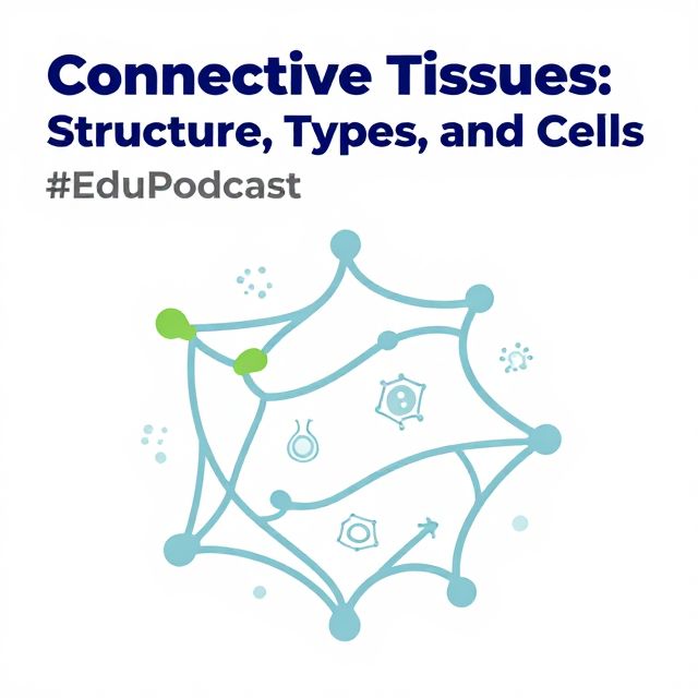
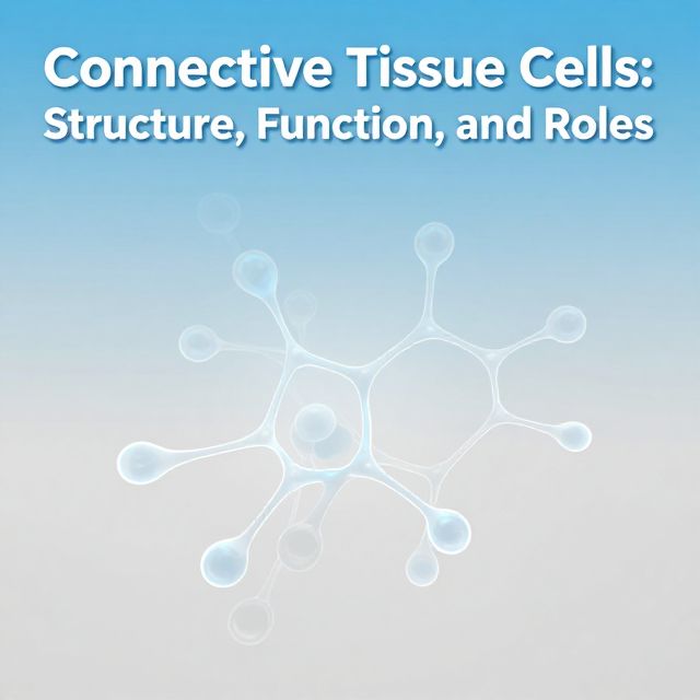
2️⃣ Connective Tissue

Connective tissue is the most diverse and abundant tissue type, found throughout the body.

📍 Locations:

  • Throughout the body, including bone, cartilage, and blood.

🎯 Functions:

  • Support and Protection: Provides structural framework and protects organs.
  • Bind Structures: Connects different tissues and organs.
  • Fill Spaces: Occupies gaps between organs.
  • Store Fat: Adipose tissue stores energy.
  • Produce Blood Cells: Bone marrow (a type of connective tissue) produces blood cells.
  • Protect Against Infection: Contains immune cells.
  • Repair Tissue Damage: Involved in wound healing.

🔑 Key Characteristics:

  • Abundant Extracellular Material (Matrix): Cells are widely spaced apart within a matrix.
  • Diverse Matrix: The matrix varies greatly depending on the tissue type.
    • 💡 Examples: Hard crystals in bone, fluid plasma in blood.

🔬 Classes of Connective Tissue:

A. Connective Tissue Proper:

  1. Loose Connective Tissue:

    • Holds organs in place and attaches epithelial tissue to underlying structures.
    • Types: Areolar tissue, reticular tissue, and adipose tissue.
    • Cells Present:
      • Cells that secrete collagen and other fibrous proteins.
      • Mast cells: Produce histamine (a blood vessel dilator) and heparin (an anticoagulant).
      • Macrophages: Immune system's first defense against invading organisms.
    • Adipose Tissue: Composed of large groups of adipose cells, each containing a droplet of fat (triglycerides) for energy storage. These can be hydrolyzed into fatty acids and secreted into the blood when energy is needed.
  2. Dense Connective Tissue:

    • Characterized by tightly packed collagen fibers, making it stronger than loose connective tissue.
    • Types:
      • Regular Dense Connective Tissue: Collagen fibers are lined up in parallel.
        • 💡 Examples: Tendons (bind muscle to bone), Ligaments (bind bone to bone).
      • Irregular Dense Connective Tissue: Collagen fibers have different orientations.
        • 💡 Examples: Covers organs (e.g., capsules of kidneys and adrenal glands), muscles, nerves, and bones.

B. Special Connective Tissues:

  • Includes cartilage, bone, and blood. (Detailed characteristics not provided in source material).

3️⃣ Muscle Tissue

Muscle tissue is specialized for contraction, enabling movement and maintaining body functions.

🎯 Functions:

  • Motion:
    • Movement of the whole body.
    • Localized movements (e.g., heart beating, digestion).
  • Maintenance of Posture: Helps hold the body upright in stationary positions.
  • Heat Production: Occurs when muscles contract, helping to maintain normal body temperature.

🔑 Key Characteristics:

  • Striated Muscles: Skeletal and cardiac muscles are called striated due to transverse stripes visible under a microscope.
  • Voluntary vs. Involuntary Control:
    • Voluntary: Contraction is consciously controlled (e.g., skeletal muscle).
    • Involuntary: Contraction is not consciously controlled (e.g., cardiac and smooth muscles).

🔬 Types of Muscle Tissue:

  1. Smooth Muscle:

    • Location: Found in the organs of the internal environment, or viscera (sometimes called visceral muscle).
    • Structure: Sheets of long, spindle-shaped cells, each containing a single nucleus.
    • Contraction:
      • In some tissues, cells contract only when stimulated by a nerve.
      • In others (e.g., gut wall), they may spontaneously initiate electrical impulses and contract, leading to slow, steady contractions. Nerves regulate, rather than cause, this activity.
  2. Skeletal Muscle:

    • Location: Attached to bones by tendons.
    • Function: Produces all movements of body parts in relation to each other.
    • Structure: Made up of numerous, very long muscle cells (muscle fibers) that lie parallel to each other. Each fiber is stimulated by a nerve fiber.
    • Control: Voluntary control; the nervous system can vary the strength of contraction.
  3. Cardiac Muscle:

    • Location: Found only in the heart.
    • Structure: Composed of smaller, interconnected striated muscle cells, each with a single nucleus.
    • Interconnections: Cells are linked by gap junctions, enabling them to form a single, functioning unit known as the myocardium.
    • Contraction: Certain cardiac muscle cells generate electrical impulses spontaneously, which spread across the gap junctions, causing all cells to contract in a coordinated manner.
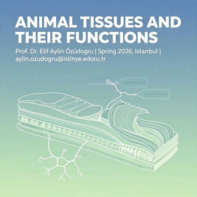
4️⃣ Nervous Tissue

Nervous tissue is the control center, integrating and coordinating all body activities.

🎯 Functions:

  • Transmit Signals (Impulses): Rapid communication across the body.
  • Coordinate, Regulate, and Integrate: Harmonizes body functions.
  • Respond to Changes: Detects and reacts to internal and external stimuli.

🔬 Cells of Nervous Tissue:

  1. Neurons:

    • Specialized to produce and conduct electrochemical impulses.
    • Structure: Each neuron consists of three main parts:
      • Cell Body (Soma): Contains the nucleus.
      • Dendrites: Thin, highly branched extensions that receive incoming stimulation and conduct electrical events towards the cell body.
      • Axon: Conducts electrical events away from the cell body.
  2. Neuroglia (Glial/Supporting Cells):

    • Do not conduct electrical impulses.
    • Functions: Support and insulate neurons, and eliminate foreign materials in and around neurons.

📊 Major Vertebrate Organ Systems

The four primary tissue types combine to form organs, which then work together in complex organ systems to maintain life. Here's an overview of the major vertebrate organ systems:

  1. Integumentary System: Protects body from injury, dehydration, pathogens; moderates temperature; excretes wastes; detects external stimuli.
  2. Nervous System: Detects external and internal stimuli; coordinates responses; integrates organ system activities.
  3. Endocrine System: Secretes hormones that control activity of other organ systems.
  4. Muscular System: Moves the body and its parts; maintains posture; produces heat.
  5. Skeletal System: Supports and protects body parts; site of muscle attachment; produces red blood cells; stores minerals.
  6. Circulatory System: Distributes materials and heat throughout the body; helps maintain pH.
  7. Lymphatic System: Collects and returns tissue fluid to the blood; defends against infection and cancers.
  8. Respiratory System: Takes in oxygen for aerobic respiration; expels carbon dioxide.
  9. Digestive System: Takes in food and water; breaks down food and absorbs nutrients; eliminates residues.
  10. Urinary System: Maintains volume and composition of blood; excretes excess fluid and wastes.
  11. Reproductive System:
    • Female: Produces eggs; nourishes and protects developing offspring.
    • Male: Produces sperm and transfers them to a female.

Kendi çalışma materyalini oluştur

PDF, YouTube videosu veya herhangi bir konuyu dakikalar içinde podcast, özet, flash kart ve quiz'e dönüştür. 1.000.000+ kullanıcı tercih ediyor.

Sıradaki Konular

Tümünü keşfet
Understanding Connective Tissues and Their Cells

Understanding Connective Tissues and Their Cells

Explore the fundamental types, structures, and cellular components of connective tissues, including their crucial roles in the body's support, protection, and immunity.

Özet 25 15
Fundamental Biological Principles and Nutrition

Fundamental Biological Principles and Nutrition

An academic summary covering biological organization from cells to organ systems, key cellular processes, major organ system functions, and essential nutritional components with their identification tests.

5 dk Özet 25 15
Understanding Animal Tissues: Structure, Function, and Development

Understanding Animal Tissues: Structure, Function, and Development

Explore the intricate world of animal tissues, focusing on epithelium and connective tissues. Learn about their embryonic origins, diverse structures, and vital functions in the body.

8 dk Özet 25 15
Connective Tissue Cells: Structure, Function, and Roles

Connective Tissue Cells: Structure, Function, and Roles

This summary provides an academic overview of connective tissue cells, detailing their classification, morphology, specific functions, and critical roles in tissue maintenance, repair, and immune responses.

9 dk Özet 25 15
Connective Tissue: Structure, Types, and Functions

Connective Tissue: Structure, Types, and Functions

An academic overview of connective tissue, detailing its embryonic origins, classifications into proper and specialized types, and the composition and roles of its fibrous and extracellular matrix components.

7 dk Özet 25 15
Fundamental Principles of Biological Organization and Nutrition

Fundamental Principles of Biological Organization and Nutrition

This summary explores the foundational concepts of biological organization, from cells to organ systems, and delves into essential nutritional components and their biochemical identification methods.

5 dk Özet 25 15
Bone Tissue and Joint Anatomy

Bone Tissue and Joint Anatomy

An academic summary detailing the composition, structure, cellular components, development, and pathologies of bone tissue, alongside an overview of joint types and synovial joint anatomy.

14 dk Özet 25 15
Cartilage and Bone Structure and Growth

Cartilage and Bone Structure and Growth

This summary provides an academic overview of cartilage structure, extracellular matrix, chondrogenesis, distinct cartilage types, and the mechanisms of bone growth and thickening.

6 dk Özet 25 15