Comprehensive Study Guide: Cellular Adaptation, Injury, and Death
Source Information: This study material has been compiled from a lecture audio transcript and copy-pasted text provided by the user. All content has been translated and organized into English.
Introduction to Cellular Responses 📚
Cells are constantly exposed to various stimuli, both physiological (normal body functions) and pathological (disease-related). Their ability to respond to these changes is crucial for survival and maintaining homeostasis. This guide explores how cells adapt to stress, the mechanisms of cellular injury when adaptation fails, and the different ways cells die. We will also cover intracellular accumulations and pathological calcification.
I. Cellular Adaptations ✅
Adaptation refers to reversible functional or structural responses of cells to physiological or pathological stimuli. These responses allow cells to survive and function in altered environments.
A. Atrophy 📉
📚 Definition: A decrease in cell and organelle size, often accompanied by a reduction in cell number.
1. Types of Atrophy
- Physiological Atrophy:
- During embryogenesis: Atrophy of the notochord and thyroglossal duct.
- Post-partum: Reduction in uterus size.
- Menopause: Atrophy of mammary glands, endometrium, and vaginal epithelium.
- Pathological Atrophy:
- Disuse Atrophy: Reduced workload (e.g., limb in a cast).
- Denervation Atrophy: Loss of nerve supply.
- Ischemic Atrophy: Reduced blood supply (most common cause).
- Inadequate Nutrition: Malnutrition.
- Loss of Endocrine Stimulation: (e.g., menopausal atrophy).
- Pressure Atrophy: Prolonged pressure on tissue.
- Senile Atrophy: Atrophy due to aging.
- Chronic Inflammation: Can lead to atrophy.
2. Pathogenesis of Atrophy
- Decreased protein synthesis.
- Increased protein degradation, primarily via the ubiquitin-proteasome pathway.
- Lipofuscin pigment (wear-and-tear pigment) accumulates in aging and atrophic cells, especially in the liver and myocardium.
3. Autophagy 💡
📚 Definition: The process by which a cell digests its own components.
- Most commonly observed in nutrient deprivation and atrophic cells.
- The phagophore membrane, involved in autophagosome formation, primarily originates from the endoplasmic reticulum (ER).
- Microtubule-associated protein light chain 3 (LC3) can be used as an autophagy marker.
- Atg genes are associated with autophagy.
B. Hypertrophy 📈
📚 Definition: An increase in the size of cells and organs.
- Often occurs alongside hyperplasia, but in non-dividing cells (e.g., skeletal and cardiac muscle), only hypertrophy is observed.
1. Types of Hypertrophy
- Physiological Hypertrophy:
- Pregnancy: Enlargement of the uterus due to myocyte hypertrophy.
- Exercise-induced hypertrophy (e.g., skeletal muscle).
- Pathological Hypertrophy:
- Most common stimulus is increased workload (e.g., cardiac hypertrophy in hypertension).
2. Pathogenesis of Hypertrophy
- Increased protein synthesis and an increase in the number of granular endoplasmic reticulum.
- PI3K/AKT pathway plays a role in physiological hypertrophy.
- G protein-coupled pathways are involved in pathological hypertrophy.
- Activation of transcription factors like GATA4, NFAT, and MEF2 increases protein synthesis.
- In cardiac hypertrophy, increased ANF gene expression is observed.
C. Hyperplasia ➕
📚 Definition: An increase in the number of cells, secondary to growth factor or hormonal stimulation.
1. Types of Hyperplasia
- Physiological Hyperplasia:
- Pregnancy: Uterine enlargement and mammary gland development.
- Puberty: Mammary gland development.
- Compensatory hyperplasia: Liver regeneration after hepatectomy, bone marrow hyperplasia after hemorrhage.
- Pathological Hyperplasia:
- Endometrial hyperplasia due to estrogen excess (can be precancerous).
- Benign prostatic hyperplasia due to androgen excess.
- Papillomatous lesions caused by HPV (can be precancerous).
2. Pathogenesis of Hyperplasia
- Growth factors and hormones stimulate the proliferation of mature cells.
- Activation of stem cells also contributes to this process.
D. Metaplasia 🔄
📚 Definition: A reversible change where one mature cell type is replaced by another mature cell type, usually in response to chronic irritation.
- Most commonly observed in epithelial tissue.
- The most frequent example is squamous metaplasia.
1. Examples of Metaplasia
- Respiratory System: Columnar epithelium transforms into squamous epithelium in smokers or due to Vitamin A deficiency/excess.
- Secretory Ducts: Presence of stones (pancreas, salivary glands, bile ducts) can cause normal secretory columnar epithelium to become stratified squamous epithelium.
- Chronic Cystitis: Transitional bladder epithelium transforms into squamous epithelium.
- Barrett's Esophagus: Squamous epithelium of the esophagus transforms into intestinal columnar epithelium, often with goblet cells (most valuable finding).
- ⚠️ Progression: Metaplasia → Dysplasia → Carcinoma (e.g., Gastroesophageal Reflux → Barrett's Metaplasia → Dysplasia → Esophageal Adenocarcinoma).
- Non-precancerous Metaplasia: Myositis ossificans (mesenchymal metaplasia) and apocrine metaplasia in the breast are generally not considered precancerous.
2. Pathogenesis of Metaplasia
- Involves the reprogramming of stem cells within the normal tissue, rather than a phenotypic change of already differentiated mature cells.
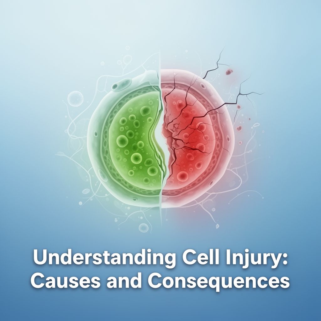
II. Cellular Injury 💥
Cellular injury occurs when cells are subjected to stress that is so severe that they are no longer able to adapt or when they are exposed to inherently damaging agents.
A. Causes and Initial Response (Hypoxia/Ischemia)
- Most common cause of clinically significant cell damage: Hypoxia.
- Most common cause of hypoxia: Ischemia due to arterial occlusion.
- Initial impact of hypoxia: Aerobic respiration is affected first.
- First organelle affected by hypoxia: Mitochondria.
- Consequence of mitochondrial dysfunction: Decreased intracellular ATP production.
1. Effects of ATP Depletion
- Na+/K+ pump failure: Leads to increased intracellular sodium.
- Cellular swelling: Sodium draws water into the cell, causing water-filled vacuoles in the cytoplasm (acute cellular swelling).
- 💡 Key Point: Cellular swelling is a widespread and reversible injury, and it is the first morphological change observed in cell injury.
- This is followed by fatty change (steatosis).
- Anaerobic glycolysis: In the absence of oxygen, cells switch to anaerobic glycolysis to produce ATP.
- This leads to a decrease in intracellular pH and a reduction in intracellular glycogen.
- The drop in pH causes chromatin clumping (chromatin condensation).
- Ribosome detachment: Decreased ATP causes ribosomes to detach from the ER, leading to ER swelling and cessation of protein synthesis.
- Increased intracellular lipid accumulation.
2. Calcium Influx
- ATP depletion impairs calcium pumps, leading to increased intracellular calcium.
- Increased intracellular calcium activates damaging enzymes: ATPases, phospholipases, proteases, and endonucleases.
- This causes phospholipid damage, cell membrane damage, breakdown of cytoskeletal proteins, and nuclear chromatin damage, ultimately leading to cell death.
- Calcium also disrupts mitochondrial permeability, increasing cytochrome c release, which initiates apoptosis.
B. Mechanisms of Injury
- Mitochondrial Damage:
- Causes: Increased calcium, reactive oxygen species (ROS), ischemia, toxins.
- Consequences:
- Increased mitochondrial permeability → impaired oxidative phosphorylation → ATP depletion → Necrosis.
- Increased ROS production → Necrosis.
- Increased outer membrane permeability → cytochrome c leakage → caspase activation → Apoptosis.
- Reactive Oxygen Species (ROS) / Oxidative Stress:
- Mechanisms of damage:
- Membrane lipid peroxidation (most important initial damage).
- Oxidative modification of proteins and enzymes.
- DNA damage.
- Key ROS products: Hydroxyl radical (OH•), superoxide anion (O2•-), hydrogen peroxide (H2O2), peroxynitrite (ONOO-).
- Cellular defense against ROS:
- Antioxidant enzymes: Superoxide dismutase, glutathione peroxidase, catalase.
- Antioxidant substances: Vitamins E and C, glutathione.
- Mechanisms of damage:
C. Ischemia-Reperfusion Injury 🩸
📚 Definition: Damage that occurs when blood flow is restored to tissues previously deprived of oxygen (ischemia), often accelerating cell death.
- Most commonly observed in the heart and brain.
- Mechanisms:
- Oxidative stress (due to sudden influx of oxygen).
- Increased intracellular calcium levels.
- Inflammation.
- Complement system activation (e.g., IgM).
D. Reversible vs. Irreversible Injury ⚠️
1. Reversible Cell Injury
- Morphological features:
- Cellular swelling (first morphological sign).
- Microvillus disruption or loss.
- Blebs on the cell membrane.
- Loosening of intercellular junctions.
- Detachment of ribosomes from the ER.
- ER swelling.
- Decreased protein synthesis.
- Cellular fatty change (steatosis).
- Mild mitochondrial swelling and small amorphous densities.
2. Irreversible Cell Injury
- Morphological features:
- Severe cell membrane damage (most important).
- Rupture of lysosomal membranes and activation of enzymes.
- Severe mitochondrial damage and large amorphous densities.
- Myelin figures (remnants of damaged phospholipid membranes).
- Nuclear changes:
- Karyopyknosis: Nuclear shrinkage and increased basophilia (also seen in apoptosis).
- Karyorrhexis: Fragmentation of the pyknotic nucleus (only in necrosis).
- Karyolysis: Fading of the nucleus due to DNA degradation by endonucleases and loss of basophilia (only in necrosis).
III. Cell Death 💀
Cell death is a fundamental process in development, tissue homeostasis, and disease. It can occur through various mechanisms.
A. Necrosis ☠️
📚 Definition: Uncontrolled cell death, typically triggered by external injury, leading to cell swelling, membrane rupture, and release of intracellular contents, often eliciting an inflammatory response.
- Most common cause of cell death: Ischemia.
- Necrotic cells stain eosinophilic and appear glassy and homogeneous. They can also undergo dystrophic calcification.
- Eosinophilic staining: Due to increased denatured globulins and loss of RNA (RNA normally stains basophilic).
- Glassy, homogeneous appearance: Due to loss of glycogen.
- Nuclear changes: Karyopyknosis, Karyorrhexis, Karyolysis (as described above).
1. Morphological Types of Necrosis
- Coagulative Necrosis:
- Most common type, typically follows ischemia (except in the brain).
- Enzyme and protein denaturation due to lactic acidosis.
- Cell outlines are preserved for several days ("ghost cells") because enzymatic degradation is inhibited.
- Example: Myocardial infarction.
- Liquefactive Necrosis:
- Characterized by enzymatic digestion of dead cells, resulting in a viscous liquid mass (pus).
- Occurs in abscesses and ischemic injury to the central nervous system (brain infarction).
- Gangrenous Necrosis:
- Refers to coagulative necrosis affecting a limb, typically the lower leg, due to ischemia.
- Dry gangrene: Pure coagulative necrosis, often seen in diabetics.
- Wet gangrene: If anaerobic bacterial infection superimposes, leading to liquefactive necrosis.
- Caseous Necrosis:
- Characteristic of tuberculosis infection, often accompanied by granulomas.
- Grossly appears cheese-like (friable, white-yellow).
- Microscopically, amorphous granular debris with fragmented cells, surrounded by a granulomatous inflammatory border.
- Fat Necrosis:
- Refers to focal areas of fat destruction.
- Traumatic fat necrosis: In breast tissue.
- Enzymatic fat necrosis: In acute pancreatitis, activated pancreatic enzymes leak into adipose tissue, breaking down triglycerides into fatty acids that combine with calcium to form chalky white deposits (saponification).
- Fibrinoid Necrosis:
- Seen in immune complex vasculitis (e.g., Polyarteritis Nodosa).
- Characterized by a pink, homogeneous, amorphous appearance in vessel walls due to deposition of immune complexes and fibrin.
B. Apoptosis 👻
📚 Definition: Programmed cell death, a highly regulated process where a cell initiates its own demise in a controlled manner, without eliciting an inflammatory response. Often called "cell suicide."
- Activated caspases digest the cell's DNA, cytoplasmic, and nuclear proteins.
- The cell breaks into small fragments called apoptotic bodies, which are then phagocytosed by macrophages.
- Receptors for phagocytosis of apoptotic bodies: Phosphatidylserine, Thrombospondin, and C1q.
- First sign of apoptosis: Cellular shrinkage and condensation.
- Most characteristic feature: Chromatin condensation and fragmentation.
- Key differences from necrosis:
- No inflammation or neutrophils (macrophages are present).
- Cell membrane and cellular contents remain intact (no rupture).
- Apoptotic cells appear as round or oval cells with dense eosinophilic cytoplasm and fragmented nuclear chromatin.
- Stain for apoptotic cells: Annexin V.
1. Types of Apoptosis
- Physiological Apoptosis:
- Programmed cell death during embryogenesis.
- Endometrial cells during menstruation.
- Ovarian follicular atresia during menopause.
- Regression of mammary glands after weaning.
- Elimination of autoreactive T cells during development.
- Pathological Apoptosis:
- DNA damage.
- Accumulation of misfolded proteins (ER stress).
- Duct obstruction in parenchymal organs (pancreas, parotid, kidney).
- T-lymphocyte mediated cell death in virus-infected cells (e.g., Councilman bodies in viral hepatitis).
- Death of cells that have completed their function in acute inflammation.
2. Caspases 🔪
- Enzymes central to apoptosis.
- Initiator Caspases: Caspase 8, 9, 10 (start the process).
- Effector (Executioner) Caspases: Caspase 3, 6 (carry out the cell's destruction).
- Caspase 3 is involved in both intrinsic and extrinsic pathways.
3. Intrinsic (Mitochondrial) Apoptosis Pathway (Most Common)
1️⃣ Increased mitochondrial membrane permeability. 2️⃣ Cytochrome c release from mitochondria into the cytoplasm. 3️⃣ Cytochrome c binds to APAF-1 (Apoptosis Activating Factor-1) to form the apoptosome. 4️⃣ Apoptosome activates Caspase 9. 5️⃣ Caspase 9 activates effector caspases (e.g., Caspase 3), leading to cell death.
- Gene families involved:
- Anti-apoptotic genes: Bcl-2, Bcl-xL, Mcl-1 (inhibit cytochrome c release).
- Pro-apoptotic genes: Bax, Bak (increase mitochondrial permeability and cytochrome c release).
- Sensor genes: Bim, Bid, Bad, Puma, Noxa (activate pro-apoptotic genes).
- SMAC/DIABLO: Mitochondrial protein that inhibits physiological inhibitors of apoptosis (IAPs), thereby promoting apoptosis.
4. Extrinsic (Death Receptor) Apoptosis Pathway
1️⃣ Activation of death receptors on the cell membrane (e.g., TNFR1, Fas/CD95). 2️⃣ Fas ligand on cytotoxic T lymphocytes binds to Fas on target cells (e.g., virus-infected cells, tumor cells, autoreactive T cells). 3️⃣ Binding leads to activation of FADD (Fas-Associated Death Domain). 4️⃣ FADD activates Caspase 8 or 10. 5️⃣ Caspase 8/10 activates effector caspases (e.g., Caspase 3/6), leading to cell death.
- Inhibitor: FLIP (binds to pro-caspase 8, inhibiting its activation).
5. Modulators of Apoptosis
- Promoters: P53, Bax, Bak, Bim, Bid, PUMA, NOXA, Cytochrome c, APAF-1, Smac/DIABLO.
- Inhibitors: Bcl-2, Bcl-xL, Mcl-x, Mdm2, Mdmx.
- Molecules for macrophage recognition:
- Apoptotic cells: Thrombospondin, Phosphatidylserine, C1q.
- Necrotic cells: DAMPs (Damage-Associated Molecular Patterns).
C. Other Forms of Cell Death
1. ER Stress 💡
📚 Definition: Apoptosis triggered by an excessive accumulation of misfolded proteins in the endoplasmic reticulum.
- Examples of diseases: Cystic fibrosis, familial hypercholesterolemia, Tay-Sachs disease, Alpha-1 antitrypsin deficiency, Creutzfeldt-Jakob disease, Alzheimer's disease.
2. Necroptosis 💥
📚 Definition: A form of programmed necrosis, sharing features of both necrosis and apoptosis, but caspase-independent.
- Initiated by receptor stimulation (e.g., TNFR).
- TNF receptor stimulation activates the RIPK1-RIPK3 complex (necrosome).
- This complex phosphorylates and activates SOR-MLKL proteins, leading to ROS formation and cell membrane damage (morphologically similar to necrosis).
3. Pyroptosis 🔥
📚 Definition: A highly inflammatory form of programmed cell death, characterized by caspase activation (Caspase 1, then 4, 5) and release of inflammatory mediators (e.g., IL-1), often leading to fever.
- Similar to apoptosis in caspase involvement, but distinct due to inflammation.
4. Ferroptosis ⚙️
📚 Definition: A type of cell death caused by excessive iron accumulation or reactive oxygen species damage, leading to the collapse of the antioxidant defense system and cell membrane damage.
- Key features: Fragmentation of the mitochondrial outer membrane and disruption of cristae structure.
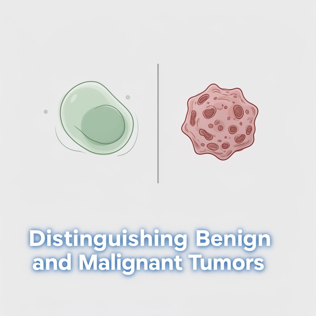
D. Comparison: Necrosis vs. Apoptosis 📊
| Feature | Apoptosis | Necrosis | | :------------------ | :------------------------------------------- | :----------------------------------------------- | | Cell Size | Shrinkage (condensation) | Swelling (enlargement) | | Nucleus | Chromatin condensation & fragmentation | Pyknosis → Karyorrhexis → Karyolysis | | Cell Membrane | Intact | Ruptured | | Cellular Contents | Intact, may form apoptotic bodies | Enzymatic digestion, leaks out of cell | | Inflammation | Absent | Often present | | Affected Cells | Single or few cells | Many cells | | Physiological/Pathological | Often physiological (elimination of unwanted cells) | Always pathological |
IV. Intracellular Accumulations 📦
Cells can accumulate various substances due to metabolic derangements, genetic defects, or exogenous overload.
A. Lipids
- Steatosis (Triglycerides):
- Most common in the liver, then myocardium, less frequently in muscle and kidneys.
- Most common causes: Alcohol and non-alcoholic fatty liver disease.
- Cholesterol:
- Accumulates in conditions like atherosclerosis, xanthomas, xanthelasmas, cholesterosis, and Niemann-Pick disease.
B. Proteins
- Reabsorption droplets: In proximal renal tubules (secondary to proteinuria).
- Russell bodies (intracellular) & Dutcher bodies (intranuclear): Accumulations of excessive immunoglobulins (ER-derived) in diseases like Multiple Myeloma.
C. Glycogen
- Stains purple-violet with Best Carmine or PAS stain.
D. Hyaline Change
- Homogeneous, pink, eosinophilic changes in intracellular or extracellular areas.
- Examples: Russell bodies, reabsorption droplets, hyaline arteriosclerosis, hyaline membranes, Mallory bodies (intermediate filament accumulation in alcoholic hepatitis).
E. Pigments 🎨
1. Exogenous Pigments
- Tattoo pigments: Phagocytosed by dermal macrophages; do not induce inflammation.
- Carbon (Coal pigment): Most common exogenous pigment.
- Causes: Smoking, air pollution, coal mining.
- Accumulation in lungs and lymph nodes is called Anthracosis.
2. Endogenous Pigments
- Lipofuscin (Lipochrome / Wear-and-Tear pigment):
- Lipid and protein mixture, accumulates due to free oxygen and toxic radicals.
- Harmless to the cell. Accumulates with age and atrophy.
- Most common in myocardium and liver.
- Melanin:
- Formed by tyrosine oxidation. The only endogenous brown-black pigment.
- Hemosiderin:
- Iron-containing, golden-yellow or brown pigment derived from hemoglobin.
- Locally seen in hemorrhage sites.
- Systemic accumulation (Hemosiderosis):
- Hemolytic anemia.
- Blood transfusions.
- Hemochromatosis.
- Bilirubin:
- Yellow-brown pigment derived from hemoglobin, found in bile.
- Homogentisic acid (Ochronosis):
- Accumulates in Alkaptonuria (homogentisic acid oxidase enzyme defect).
- Causes black discoloration of skin, connective tissue, and cartilage.
V. Pathological Calcification 🦴
Abnormal deposition of calcium salts in tissues.
A. Dystrophic Calcification
📚 Definition: Calcium deposition in damaged, necrotic, or dying tissues despite normal serum calcium levels.
- Examples:
- Atheromas in atherosclerosis.
- Damaged heart valves in the elderly.
- Tuberculosis-infected lymph nodes (scrofula).
- Psammoma bodies: Lamellar calcifications seen in some carcinomas (e.g., papillary thyroid carcinoma, serous ovarian tumors, meningioma, prolactinoma, papillary renal cell carcinoma).
- Asbestos bodies.
B. Metastatic Calcification
📚 Definition: Calcium deposition in normal tissues due to hypercalcemia (elevated serum calcium levels).
- Can occur in many locations, but typically affects gastric mucosa, kidneys, lungs, systemic arteries, and pulmonary veins.
- Most typical example: Nephrocalcinosis.
- Causes of hypercalcemia:
- Increased PTH production: Primary hyperparathyroidism (adenoma/hyperplasia), secondary hyperparathyroidism (renal failure), ectopic PTH production by tumors (e.g., squamous cell carcinoma of the lung).
- Vitamin D-related diseases: Sarcoidosis (granulomas produce excess Vitamin D), Williams Syndrome (idiopathic hypercalcemia), Vitamin D intoxication.
- Excessive bone destruction: Multiple myeloma, metastases, immobilization, Paget's disease.
VI. Cellular Aging ⏳
Cellular aging is a complex process involving various mechanisms that lead to a decline in cellular function and viability over time.
- Calorie restriction: Known to extend lifespan, primarily by increasing sirtuins and inhibiting the IGF-1 signaling pathway.
- Sirtuins: Proteins that slow metabolic activity, reduce apoptosis, stimulate protein folding, and activate DNA repair genes. They are considered lifespan-extending proteins.
- Mechanisms of cellular aging:
- Reduced cell replication: Linked to telomere shortening and telomerase activity.
- Telomerase: Enzyme that maintains telomere length. Germ cells and stem cells have high telomerase activity.
- Werner Syndrome: A progeroid syndrome caused by a defect in DNA helicase, leading to accelerated aging.
- DNA damage: Accumulation of unrepaired DNA damage.
- Defective protein homeostasis: Impaired protein folding and degradation.
- Dysregulation of cell nutrient sensing.
- Reduced cell replication: Linked to telomere shortening and telomerase activity.
VII. Important Stains in Pathology 🔬
| Stain | Substance Detected | | :-------------------- | :----------------------------------------------------- | | Prussian Blue | Iron, Hemosiderin | | Masson-Fontana | Melanin | | Masson's Trichrome | Collagen (blue/green), Muscle (red), Keratin (red) | | Sudan Black / Oil Red O | Lipids, Fat | | Luxol Fast Blue | Myelin | | Orcein-Rhodamine | Copper | | PAS (Periodic Acid-Schiff) | Glycogen, Glycoproteins (mucus), Basement membranes, Fungi | | Alcian Blue | Mucin | | Congo Red | Amyloid (apple-green birefringence under polarized light) | | Warthin-Starry / Modified Giemsa | H. pylori | | Toluidine Blue / Giemsa | Mast cells (metachromatic staining) | | Phosphotungstic Acid Hematoxylin (PTAH) | Mitochondria |