CNS Membranes, CSF, Basal Ganglia, and Diencephalon - kapak
Bilim#meninges#dura mater#arachnoidea mater#pia mater

CNS Membranes, CSF, Basal Ganglia, and Diencephalon

An academic overview of the central nervous system's protective membranes, cerebrospinal fluid circulation, the basal ganglia's motor functions, and the diencephalon's sensory and regulatory roles.

juliamedicFebruary 11, 2026 ~32 dk toplam
01

Sesli Özet

12 dakika

Konuyu otobüste, koşarken, yolda dinleyerek öğren.

Sesli Özet

CNS Membranes, CSF, Basal Ganglia, and Diencephalon

0:0011:42
02

Flash Kartlar

25 kart

Karta tıklayarak çevir. ← → ile gez, ⎵ ile çevir.

1 / 25
Tüm kartları metin olarak gör
  1. 1. What are the three main layers of the meninges protecting the central nervous system, from superficial to deep?

    The three main layers of the meninges are the dura mater, arachnoidea mater, and pia mater. These membranes collectively protect the brain and spinal cord, providing a physical barrier against injury and supporting the neural tissue. They also help to maintain the chemical environment necessary for proper CNS function.

  2. 2. Describe the key characteristics and layers of the dura mater around the encephalon versus the medulla spinalis.

    Around the encephalon, the dura mater's two layers are typically fused, forming dural sinuses where they separate. In contrast, around the medulla spinalis, these two dural layers are distinct, creating the epidural space (cavum epidurale) which contains the internal vertebral venous plexus. The dura mater is a thick, inelastic structure providing robust protection.

  3. 3. Name three significant dural folds found within the cranium and state their primary function.

    Three significant dural folds are the falx cerebri, which separates the cerebral hemispheres; the falx cerebelli, located between the cerebellar hemispheres; and the tentorium cerebelli, which divides the cerebrum from the cerebellum. These folds help to compartmentalize the brain and restrict excessive movement within the cranial cavity.

  4. 4. Which artery is the largest supplier to the dura mater and why is it clinically important?

    The middle meningeal artery, a branch of the maxillary artery, is the largest artery supplying the dura mater. It is clinically important because its location within the sulcus of the temporal and parietal bones makes it vulnerable to rupture during head fractures, often leading to epidural hematomas. These hematomas typically appear biconvex on CT scans.

  5. 5. What is the leptomeninx, and what is the significance of the subarachnoid space?

    The leptomeninx refers to the combined arachnoidea mater and pia mater. The subarachnoid space, located between the arachnoidea and pia mater, is a true anatomical space, unlike the potential epidural and subdural spaces. This space contains cerebrospinal fluid (CSF) and major blood vessels, playing a crucial role in CSF circulation and nutrient exchange.

  6. 6. At what vertebral levels do the pia mater and the dura mater/arachnoidea mater terminate?

    The pia mater extends to the L1-L2 vertebral level, closely adhering to the spinal cord. The dura mater and arachnoidea mater, however, extend further down to the S2 vertebral level. This difference in termination creates a lumbar cistern, an important site for lumbar punctures to collect CSF.

  7. 7. What is the primary site of cerebrospinal fluid (CSF) production and what structures are involved?

    Cerebrospinal fluid (CSF) is primarily produced by the plexus choroideus. This structure consists of specialized ependymal cells and pia mater, located in the temporal and central parts of the lateral ventricles, the roof of the third ventricle, and the posterior wall of the fourth ventricle. The plexus choroideus actively filters blood to produce CSF.

  8. 8. How much CSF is produced daily, and what is the total volume typically present within the CNS at any given time?

    Approximately 500 milliliters of CSF are produced daily. However, the total volume within the CNS at any given time is about 150 milliliters, with 15 to 40 milliliters residing within the ventricles. This continuous production and absorption ensure a constant turnover and fresh supply of CSF.

  9. 9. What are the main structures responsible for CSF absorption, and where do they drain?

    CSF absorption is primarily facilitated by the villi arachnoideales, which are critical for this process. These villi extend through the subdural space, pierce the dura mater, and drain into the superior sagittal sinus. Clusters of these villi are known as granulationes arachnoidales or Pacchioni corpuscles, effectively returning CSF to the venous circulation.

  10. 10. Describe the path of CSF circulation from the lateral ventricles to the subarachnoid space.

    CSF flows from the two lateral ventricles into the third ventricle via the interventricular foramina (of Monro). From the third ventricle, it passes through the cerebral aqueduct (of Sylvius) into the fourth ventricle. Finally, from the fourth ventricle, CSF exits into the subarachnoid space through the two lateral foramina of Luschka and the single median foramen of Magendie.

  11. 11. Name three significant cisterns of the subarachnoid space and one key structure found within each.

    The cisterna magna (cerebellomedullary cisterna) is the largest. The quadrigeminal cisterna contains the great cerebral vein, pineal body, and posterior cerebral arteries. The cisterna interpeduncularis houses the Circle of Willis and the oculomotor nerve. These cisterns are enlargements of the subarachnoid space, allowing for CSF accumulation and passage of neurovascular structures.

  12. 12. List the main components of the basal ganglia.

    The main components of the basal ganglia (nuclei basales) include the claustrum, nucleus caudatus, nucleus lentiformis (comprising the putamen and globus pallidus), the substantia nigra, and the nucleus subthalamicus. These subcortical gray matter structures are crucial for motor control.

  13. 13. What is the striatum, and what is its role within the basal ganglia?

    The striatum, also known as the neostriatum, is formed by the nucleus caudatus and the putamen. It serves as the primary afferent (input) center for the basal ganglia. The striatum receives extensive input from the cerebral cortex and other brain regions, initiating the processing of motor commands within the basal ganglia circuit.

  14. 14. Explain the primary function of the basal ganglia in motor control.

    The basal ganglia are integral to the extrapyramidal system, playing a crucial role in the execution of repetitive, automatic movements such as driving, writing, cycling, or walking. They take over control of these movements from the frontal cortex once they are sufficiently learned, allowing the cortex to engage in other tasks. Movements learned by the basal ganglia are typically not forgotten.

  15. 15. How does Parkinson's disease relate to the basal ganglia, and what are its characteristic symptoms?

    Parkinson's disease is characterized by the loss of dopaminergic neurons in the substantia nigra pars compacta, leading to a dopamine deficiency in the neostriatum. This results in decreased motor activity in the cerebral cortex, manifesting as akinesia (difficulty initiating movement), rigidity, rest tremor, a flexor posture, and a mask-like facial expression.

  16. 16. What are the four main components of the diencephalon?

    The diencephalon comprises four key components: the thalamus, hypothalamus, epithalamus, and subthalamus. This region develops around the third ventricle and serves as a critical relay and control center for various bodily functions, but it does not directly connect to the medulla spinalis.

  17. 17. Describe the primary role of the thalamus in sensory processing.

    The thalamus is the largest part of the diencephalon and serves as a crucial relay and integration center for all sensory impulses except olfaction. It acts as a filter, transmitting only suprathreshold signals to the cerebral cortex. This ensures that only relevant sensory information reaches conscious perception, preventing sensory overload.

  18. 18. Besides sensory relay, what other functions does the thalamus contribute to?

    Beyond sensory relay, the thalamus has limbic connections, influencing emotion, learning, and memory. It also plays a role in involuntary movements through its connections with the basal ganglia. Damage to the thalamus can lead to a variety of neurological deficits, including personality changes and emotional disturbances.

  19. 19. Where is the hypothalamus located, and what is its overarching role in the body?

    The hypothalamus is located in the inferior-anterior part of the third ventricle. It is considered the "brain of the autonomic and endocrine systems" due to its overarching role in regulating metabolism and maintaining homeostasis. It integrates neural and hormonal signals to control vital bodily functions.

  20. 20. Name two specific hypothalamic nuclei and the hormones they secrete or functions they regulate.

    The supraoptic nucleus secretes vasopressin (antidiuretic hormone), which regulates water balance. The paraventricular nucleus secretes oxytocin, involved in social bonding and uterine contractions. Other nuclei regulate hunger (lateral hypothalamic nucleus), satiety (ventromedial nucleus), and circadian rhythms (suprachiasmatic nucleus).

  21. 21. How does the hypothalamus regulate body temperature?

    The hypothalamus regulates body temperature through distinct regions. The anterior hypothalamic nucleus is associated with decreasing body temperature, primarily through mechanisms like sweating and vasodilation. Conversely, the posterior hypothalamic nucleus is responsible for increasing body temperature, often by initiating shivering and vasoconstriction.

  22. 22. What structures are included in the epithalamus, and what is the function of the pineal gland?

    The epithalamus includes the habenula, commissura posterior, pretectal area, and the pineal gland. The pineal gland (glandula pinealis) secretes serotonin and melatonin. Melatonin is crucial for regulating circadian rhythms and inhibits gonadotropins, influencing the activity of other endocrine glands. Its activity is inhibited by light exposure to the retina.

  23. 23. What is the habenula's role within the epithalamus?

    The habenula, part of the epithalamus, is considered a component of the limbic system. It is involved in emotion, behavior, and the regulation of visceral and endocrine functions. It acts as a relay station, connecting the limbic forebrain with the midbrain, influencing reward and aversion pathways.

  24. 24. What is the significance of the nucleus subthalamicus within the subthalamus?

    The nucleus subthalamicus, located within the subthalamus, is considered an upward extension of the substantia nigra. It plays a crucial role in regulating muscle tone, largely independently of the cerebral cortex. Dysfunction of this nucleus can lead to involuntary movements, such as hemiballismus.

  25. 25. What are the two main components of the metathalamus and their respective functions?

    The metathalamus includes the corpus geniculatum laterale and the corpus geniculatum mediale. The corpus geniculatum laterale is where most optic tract fibers terminate, playing a role in visual processing and light reflexes. The corpus geniculatum mediale is an intermediate center in the auditory pathways, relaying auditory information to the cerebral cortex.

03

Bilgini Test Et

15 soru

Çoktan seçmeli sorularla öğrendiklerini ölç. Cevap + açıklama.

Soru 1 / 15Skor: 0

Which of the following is the correct order of meningeal layers from superficial to deep?

04

Detaylı Özet

10 dk okuma

Tüm konuyu derinlemesine, başlık başlık.

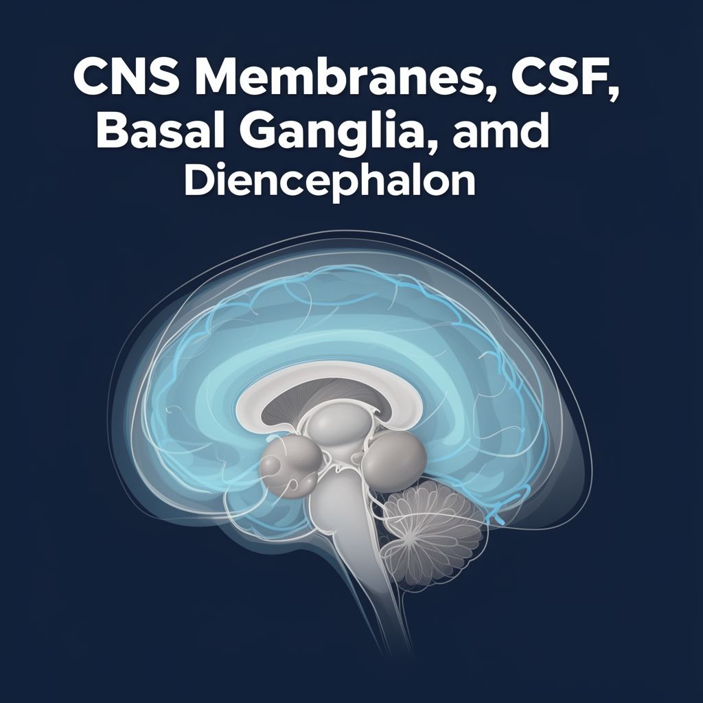
Study Material: Central Nervous System Protection and Regulation

Source Information: This study material has been compiled from a combination of copy-pasted text and a lecture audio transcript, integrating information on Central Nervous System (CNS) membranes, Cerebrospinal Fluid (CSF) circulation, Basal Ganglia, and the Diencephalon.


Introduction to CNS Protection and Regulation 🧠

The Central Nervous System (CNS), comprising the brain and spinal cord, is a highly delicate and vital organ system. It is meticulously protected by a series of membranes called meninges and cushioned by Cerebrospinal Fluid (CSF). Beyond protection, the CNS also houses critical structures like the basal ganglia for motor control and the diencephalon for sensory processing, autonomic regulation, and endocrine functions. This guide will explore these essential components, their structures, functions, and clinical significance.


I. Central Nervous System Membranes (Meninges) 📚

The meninges are three protective layers surrounding the brain and spinal cord, arranged from superficial to deep:

  • Dura Mater (Pachymeninx)
  • Arachnoidea Mater
  • Pia Mater

A. Dura Mater (Tough Mother)

The dura mater is a thick, inelastic outermost layer. It consists of two layers:

  1. Outer Endosteal Layer: Adjacent to the cranial bones, forming the inner periosteum of the skull.
  2. Inner Meningeal-Encephalic Layer: The main protective layer.

Structural Variations:

  • Around the Encephalon (Brain): The two dural layers are typically fused, separating only to form dural venous sinuses.
  • Around the Medulla Spinalis (Spinal Cord): The two dural layers are separate, creating the epidural space (cavum epidurale). This space contains the internal vertebral venous plexus.

Dural Folds (Dura Encephali): These are invaginations of the meningeal layer that divide the cranial cavity and support brain structures.

  • Falx Cerebri: Separates the two cerebral hemispheres.
  • Falx Cerebelli: Separates the two cerebellar hemispheres.
  • Tentorium Cerebelli: Divides the cerebrum from the cerebellum.
  • Cavum Trigeminale (Meckel's Cave): Encloses the trigeminal ganglion.
  • Diaphragma Sellae: Covers the hypophysis (pituitary gland).

Sensory Innervation of Dura Mater: The dura mater receives sensory innervation from various cranial and spinal nerves:

  • Anterior Cranial Fossa: Ophthalmic nerve (CN V1), Maxillary nerve (CN V2).
  • Middle Cranial Fossa: Maxillary nerve (CN V2), Mandibular nerve (CN V3).
  • Posterior Cranial Fossa: Cervical spinal nerves C2 and C3.
  • Tentorium Cerebelli & Falx Cerebri: Primarily innervated by the ophthalmic nerve (CN V1).

Arterial Supply of Dura Mater:

  • Anterior Meningeal Artery: Branch of the internal carotid artery or its ophthalmic branch.
  • Middle Meningeal Artery: The largest artery supplying the dura mater, originating from the maxillary artery.
  • Posterior Meningeal Artery: Branches from the ascending pharyngeal, occipital, or vertebral arteries.

Clinical Significance: Epidural Hematoma ⚠️

  • The Middle Meningeal Artery is clinically very important. It lies within a sulcus in the temporal and parietal bones.
  • Cause: Fractures of the temporal-parietal bone can rupture this artery, leading to bleeding into the epidural space.
  • Diagnosis: Typically appears as a biconvex (lens-shaped) image on a CT scan.
  • Note: The middle meningeal artery is projected at the Pterion, a weak point of the skull.

B. Arachnoidea Mater & Pia Mater (Leptomeninx)

Together, the arachnoidea mater and pia mater are referred to as the leptomeninx.

  • Arachnoidea Mater: A delicate, avascular membrane beneath the dura.
  • Pia Mater: The innermost, highly vascular membrane that adheres closely to the brain and spinal cord tissue, following all its contours and entering sulci.

Meningeal Spaces:

  • Epidural Space: A potential space between the dura and skull (or vertebral canal).
  • Subdural Space: A potential space between the dura and arachnoidea.
  • Subarachnoid Space: A true anatomical space between the arachnoidea and pia mater, filled with CSF.

Termination Levels of Meninges:

  • Pia Mater: Terminates at the L1-L2 vertebral level (conus medullaris).
  • Dura Mater & Arachnoidea Mater: Extend down to the S2 vertebral level.

Filum Terminale:

  • Filum Terminale Internum: An extension of the pia mater, continuing within the CSF down to S2.
  • Filum Terminale Externum: Beyond S2, it consists of pia, arachnoid, and dura, fusing to form the coccygeal ligament (lig. coccygeum).

II. Cerebrospinal Fluid (CSF) Circulation 💧

CSF is a clear, colorless fluid that surrounds the brain and spinal cord, providing protection, buoyancy, and nutrient exchange.

A. CSF Production

  • Primary Site: Choroid Plexus
  • Composition: The choroid plexus consists of specialized ependymal cells and blood vessels covered by pia mater.
  • Locations:
    • Temporal and central parts of the lateral ventricles.
    • Roof of the third ventricle.
    • Posterior wall of the fourth ventricle.
  • Production Rate: Approximately 500 mL of CSF is produced daily.
  • Total Volume: The total volume of CSF within the CNS at any given time is about 150 mL, with 15-40 mL residing in the ventricles.

B. CSF Absorption

  • Most Important Pathway: Arachnoid Villi (also known as Pacchionian Corpuscles or Arachnoid Granulations when clustered).
    • These villi extend through the subdural space, pierce the dura, and drain CSF into the Superior Sagittal Sinus.
  • Other Pathways:
    • Perineural spaces of cranial nerves.
    • Direct venous openings.
    • Perilymph of the inner ear.

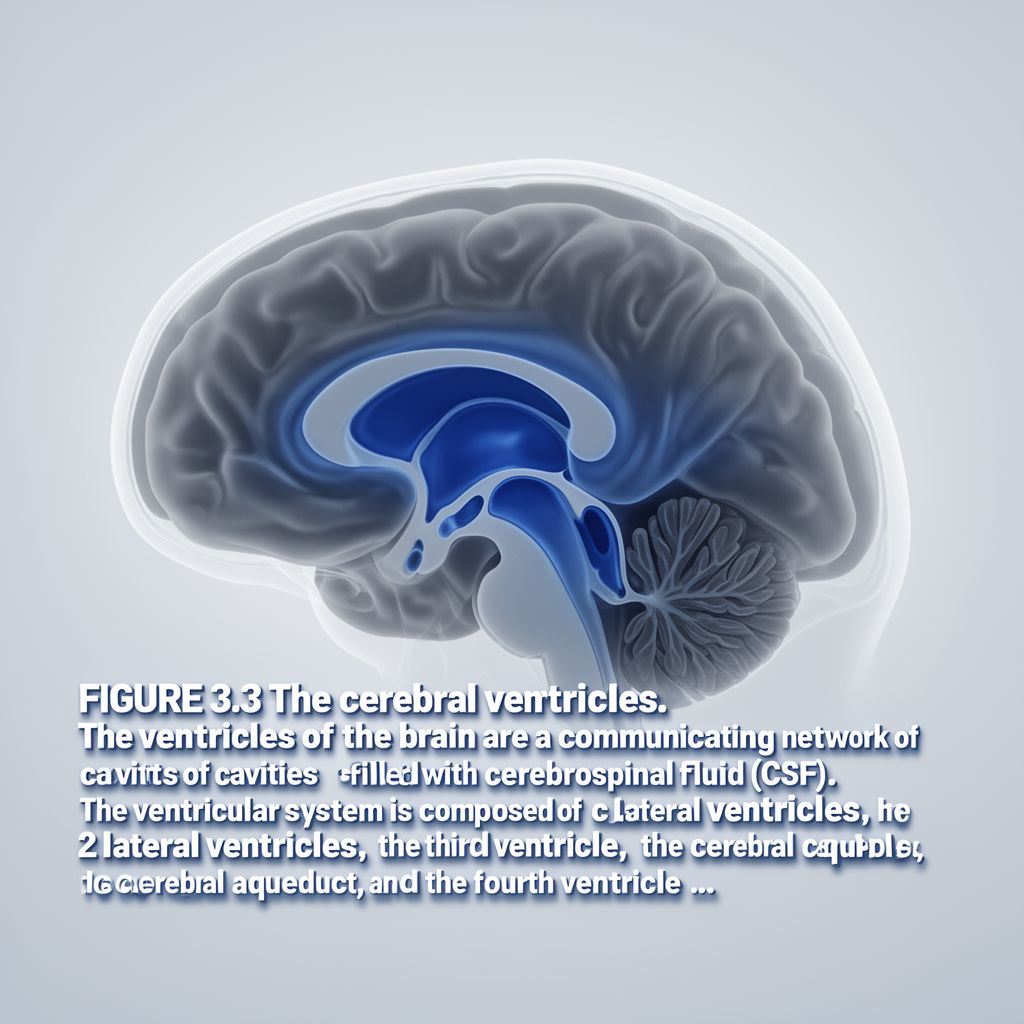
C. Ventricular System

The CSF circulates through a series of interconnected cavities within the brain.

  1. Lateral Ventricles (Two):

    • Each has four parts: central part, frontal horn, occipital horn, temporal horn.
    • Separated by the septum pellucidum, which connects superiorly to the corpus callosum and inferiorly to the fornix.
    • CSF flows from lateral ventricles to the third ventricle via the interventricular foramina (of Monro).
  2. Third Ventricle (One):

    • Located between the two halves of the diencephalon.
    • Contains several recesses: pineal, suprapineal, infundibular, supraoptic recesses.
    • CSF flows from the third ventricle to the fourth ventricle via the cerebral aqueduct (of Sylvius).
  3. Fourth Ventricle (One):

    • Located between the medulla oblongata, pons, and cerebellum.
    • Roof: Formed by the superior cerebellar peduncle and superior medullary velum.
    • Floor: Formed by the inferior cerebellar peduncle and inferior medullary velum (rhomboid fossa).
    • CSF exits the fourth ventricle into the subarachnoid space through:
      • Two lateral foramina (of Luschka)
      • One median foramen (of Magendie)

D. CSF Flow and Cisterns

After exiting the fourth ventricle, CSF enters the subarachnoid space, which contains enlargements called cisterns.

  • Cisterna Magna (Cerebellomedullary Cisterna):
    • The largest cisterna.
    • Located posterior to the medulla oblongata and inferior to the cerebellum.
  • Quadrigeminal Cisterna:
    • Contains the great cerebral vein, pineal body, and posterior cerebral arteries.
  • Chiasmatic Cisterna:
    • Surrounds the optic chiasm and hypophysis.
  • Interpeduncular Cisterna:
    • Houses the Circle of Willis and the oculomotor nerve (CN III).
  • Lateral Fossa Cisterna:
    • Contains the middle cerebral artery.
  • Pontocerebellar Cisterna (Prepontine Cisterna):
    • Associated with the basilar artery and the abducens nerve (CN VI).

III. Basal Ganglia (Nuclei Basales) 📊

The basal ganglia are a group of subcortical gray matter nuclei located deep within the telencephalon, crucial for motor control.

A. Components

The main components include:

  • Claustrum
  • Nucleus Caudatus
  • Nucleus Lentiformis: Composed of the Putamen and Globus Pallidus.
  • Substantia Nigra (located in the midbrain, functionally associated)
  • Nucleus Subthalamicus (located in the diencephalon, functionally associated)

Key Groupings:

  • Striatum (Neostriatum): Formed by the Caudate Nucleus + Putamen. This is the primary afferent (input) center of the basal ganglia.
  • Corpus Striatum: Formed by the Caudate Nucleus + Lentiform Nucleus.
  • Globus Pallidus: The primary efferent (output) center of the basal ganglia.

B. Function: Extrapyramidal System

The basal ganglia are integral to the extrapyramidal system, which is responsible for:

  • Automatic, Repetitive Movements: Such as driving, writing, cycling, or walking.
  • Motor Learning: When a movement is repeated frequently, its control shifts from the frontal cortex (e.g., Area 6) to the basal ganglia. This allows the cerebral cortex to focus on other tasks.
  • Unforgettable Movements: Movements learned by the basal ganglia are typically not forgotten.
  • Movement Characteristics: Basal ganglia-controlled movements are often characteristic, fast, and subcortical. Cortical movements tend to be slower, goal-oriented, and less automatic.

Pathways:

  • Direct Pathway: Generally excites the motor cortex.
  • Indirect Pathway: Generally inhibits the motor cortex.

C. Clinical Significance: Parkinson's Disease ⚠️

  • Cause: Loss of dopaminergic neurons in the substantia nigra pars compacta.
  • Mechanism: Leads to dopamine deficiency in the neostriatum (striatum).
  • Result: Decreased motor activity in the cerebral cortex.
  • Symptoms:
    • Akinesia: Difficulty initiating movement.
    • Rigidity: Increased muscle tone.
    • Rest Tremor: Tremor that occurs at rest.
    • Flexor Posture: Stooped posture.
    • Mask-like Face: Reduced facial expressions.
    • Difficulty with complex movements.
  • Treatment: Often involves L-DOPA to replenish dopamine.
  • Note: Damage to basal ganglia can cause involuntary, repetitive movements, often observed at rest, which may disappear when the cortex is actively engaged.

IV. Diencephalon 💡

The diencephalon is a central part of the brain, located between the mesencephalon and the cerebral hemispheres. All its components develop around the third ventricle and do not directly connect to the spinal cord.

A. Thalamus

The thalamus is the largest part of the diencephalon and a crucial relay and integration center.

  • Function:
    • Sensory Relay: Receives and integrates all sensory impulses except olfaction, filtering them and sending only suprathreshold signals to the cerebral cortex.
    • Limbic System Connections: Involved in emotion, learning, and memory.
    • Motor Control: Through connections with the basal ganglia, it plays a role in involuntary movements.
  • Key Structures:
    • Pulvinar Thalami: Posterior end of the thalamus, extending towards the superior colliculus; involved in visual processing.
    • Stratum Zonale: White matter layer covering the superior thalamic surface.
    • Internal Medullary Lamina: White matter dividing the thalamus into three main nuclear groups.
    • Interthalamic Adhesion (Massa Intermedia): A gray matter mass connecting the two thalami (present in about 70% of individuals).
    • Medial & Lateral Geniculate Bodies: Part of the metathalamus, functionally associated with the thalamus (see Metathalamus section).
  • Thalamic Nuclei (Examples):
    • Anterior Thalamic Nuclei: Emotion, memory, learning, attention.
    • Mediodorsal Nucleus (Medial Group): Mental behavior, memory, limbic system, prefrontal connections.
    • Lateral Dorsal Nucleus (Lateral Group): Limbic functions.
    • Ventral Anterior & Ventral Lateral Nuclei (Ventral Group): Influence motor cortex.
    • Ventral Posteromedial Nucleus: Receives sensory input from cranial nerves.
    • Ventral Posterolateral Nucleus: Receives sensory input from spinal nerves.
  • Arterial Supply: Primarily from the Posterior Cerebral Artery.
  • Thalamic Lesions: Often arterial, can cause contralateral anesthesia, personality changes, emotional disturbances, and amnesia.

B. Hypothalamus

Located in the inferior-anterior part of the third ventricle, the hypothalamus is considered the "brain" of the Autonomic Nervous System (ANS) and Endocrine System.

  • Function:
    • Homeostasis: Regulates metabolism and maintains internal balance.
    • ANS Control: Manages the sympathetic and parasympathetic divisions.
      • Anterior/Preoptic Area: Parasympathetic functions.
      • Posterior/Lateral Parts: Sympathetic functions.
    • Endocrine Control: Directly influences the pituitary gland.
  • Key Structures & Nuclei:
    • Optic Chiasm
    • Tuber Cinereum: A bulge in the floor of the third ventricle.
    • Mammillary Bodies
    • Preoptic Area: Structurally telencephalic, but functionally hypothalamic.
    • Median Eminence: Along with the infundibulum and posterior pituitary, forms the neurohypophysis.
    • Supraoptic Nucleus: Secretes Vasopressin (ADH).
    • Paraventricular Nucleus: Secretes Oxytocin.
    • Lateral Hypothalamic Nucleus: Signals hunger.
    • Ventromedial Hypothalamic Nucleus: Signals satiety.
    • Suprachiasmatic Nucleus: Regulates circadian rhythms.
    • Anterior Hypothalamic Nucleus: Decreases body temperature.
    • Posterior Hypothalamic Nucleus: Increases body temperature.

C. Epithalamus

The epithalamus forms the posterior border of the diencephalon.

  • Components:
    • Habenula: Part of the limbic system, involved in emotion, behavior, and regulation of visceral and endocrine functions (e.g., salivation, gastrointestinal motility).
    • Posterior Commissure
    • Pretectal Area
    • Pineal Gland (Glandula Pinealis):
      • Secretes Serotonin and Melatonin.
      • Light exposure to the retina inhibits pineal gland activity (e.g., eye masks for jet lag).
      • Melatonin inhibits gonadotropins and influences other endocrine glands (pituitary, pancreas, adrenal, parathyroid, gonads), generally causing decreased activity.
      • With age, it often calcifies, forming Acervulus Cerebri (brain sand), visible on X-rays.

D. Subthalamus

  • Nucleus Subthalamicus:
    • Considered an upward extension of the substantia nigra.
    • Functionally associated with the basal ganglia.
    • Regulates muscle tone independently of the cortex.

E. Metathalamus

The metathalamus is involved in primary reflex responses to light and sound.

  • Corpus Geniculatum Laterale (Lateral Geniculate Body - LGN):
    • Termination site for most optic tract fibers.
    • Involved in visual processing and light reflexes (fibers pass via the brachium of superior colliculus to the pretectal area for light reflex without synapsing in LGN).
  • Corpus Geniculatum Mediale (Medial Geniculate Body - MGN):
    • An intermediate center in auditory pathways.

Conclusion: Integrated Functions of CNS Protective and Regulatory Systems ✅

The CNS is a marvel of biological engineering, with intricate protective layers and fluid systems safeguarding its delicate neural tissue. The meninges and CSF provide mechanical protection, buoyancy, and a stable chemical environment. Simultaneously, the basal ganglia ensure the smooth execution of learned motor skills, while the diencephalon acts as a central hub for sensory integration, autonomic control, and endocrine regulation. These interconnected systems highlight the complex organization essential for maintaining neurological health and overall bodily homeostasis.

Kendi çalışma materyalini oluştur

PDF, YouTube videosu veya herhangi bir konuyu dakikalar içinde podcast, özet, flash kart ve quiz'e dönüştür. 1.000.000+ kullanıcı tercih ediyor.

Sıradaki Konular

Tümünü keşfet
Anatomy of the Nervous System

Anatomy of the Nervous System

An academic overview of the nervous system, detailing the Central Nervous System (CNS) and Peripheral Nervous System (PNS), their components, functions, and associated disorders.

8 dk Özet 25 15
Human Brain Anatomy: Ventricles, Terminology, and Structures

Human Brain Anatomy: Ventricles, Terminology, and Structures

Explore the intricate anatomy of the human brain, covering the ventricular system, essential directional terms, embryonic development, and the major divisions and structures of the adult brain.

25 15
Understanding the Borehole Environment

Understanding the Borehole Environment

This summary provides an academic overview of the borehole environment, detailing its characteristics, influencing factors, and significance in subsurface investigations and resource extraction.

6 dk Özet 15
Types of Dissolution and Solution Concentration

Types of Dissolution and Solution Concentration

Explore the different ways substances dissolve, including physical and chemical dissolution, and understand key concentration units like molarity and parts per million (ppm).

Özet 15 Görsel
The Musculoskeletal System: Structure, Function, and Locomotion

The Musculoskeletal System: Structure, Function, and Locomotion

Explore the intricate musculoskeletal system, its components, functions, and the mechanisms of locomotion in various organisms, with a detailed focus on the human body.

Özet 25 15 Görsel
The Nervous and Endocrine Systems: Body's Control Centers

The Nervous and Endocrine Systems: Body's Control Centers

Explore the intricate workings of the nervous and endocrine systems, their structures, functions, and how they maintain the body's homeostasis.

Özet 25 15 Görsel
The Reproductive System: Cell Division and Reproduction

The Reproductive System: Cell Division and Reproduction

Explore the fundamental processes of cell division and the diverse strategies of reproduction, including asexual and sexual methods, gametogenesis, fertilization, and the human reproductive systems.

Özet 25 15 Görsel
Introduction to Radioactivity and Its Applications

Introduction to Radioactivity and Its Applications

This summary provides an academic overview of radioactivity, covering fundamental concepts, types of radiation, decay processes, biological effects, detection methods, and diverse applications in medicine, industry, and dating.

8 dk Özet 25 15