Anatomy of the Nervous System - kapak
Bilim#nervous system#central nervous system#peripheral nervous system#brain

Anatomy of the Nervous System

An academic overview of the nervous system, detailing the Central Nervous System (CNS) and Peripheral Nervous System (PNS), their components, functions, and associated disorders.

aleyblgnMarch 29, 2026 ~24 dk toplam
01

Sesli Özet

8 dakika

Konuyu otobüste, koşarken, yolda dinleyerek öğren.

Sesli Özet

Anatomy of the Nervous System

0:008:07
02

Flash Kartlar

25 kart

Karta tıklayarak çevir. ← → ile gez, ⎵ ile çevir.

1 / 25
Tüm kartları metin olarak gör
  1. 1. What are the two primary divisions of the nervous system?

    The nervous system is fundamentally divided into two main components: the Central Nervous System (CNS) and the Peripheral Nervous System (PNS). The CNS acts as the central processing unit, while the PNS serves as the communication link between the CNS and the rest of the body, transmitting information.

  2. 2. What structures constitute the Central Nervous System (CNS)?

    The Central Nervous System (CNS) is composed of the brain and the spinal cord. These two structures work together to integrate and process information, serving as the command and control center for the entire body, orchestrating complex functions.

  3. 3. What structures make up the Peripheral Nervous System (PNS)?

    The Peripheral Nervous System (PNS) consists of nerves and ganglia. Nerves are bundles of axons that transmit signals, while ganglia are clusters of nerve cell bodies located outside the CNS. The PNS is responsible for transmitting information to and from the CNS.

  4. 4. Differentiate between afferent and efferent neurons in the PNS.

    Afferent neurons, also known as sensory neurons, transmit sensory input from the body's periphery (e.g., skin, organs) to the CNS for processing. Efferent neurons, or motor neurons, carry motor output commands from the CNS to various effector organs like muscles and glands, initiating responses.

  5. 5. List at least three sources of sensory input conveyed by the PNS.

    Sensory inputs conveyed by the PNS originate from diverse sources such as the nose (smell), eyes (sight), ears (hearing), taste buds (taste), skin (touch, temperature, pain), muscles, joints (proprioception), and internal organs (visceral sensations). These inputs provide the CNS with information about the internal and external environment.

  6. 6. What are the four principal regions into which the brain is organized?

    The brain is organized into four principal regions: the Cerebrum, the Diencephalon, the Brainstem, and the Cerebellum. Each region has specialized functions that contribute to the overall complexity and capabilities of the brain, from higher-order thinking to vital life support.

  7. 7. Describe the main components and a key function of the Cerebrum.

    The Cerebrum features an outer layer of gray matter known as the cerebral cortex and contains deep subcortical nuclei. These include the basal nuclei, crucial for cognitive processing and the planning of movements, and the basal forebrain, important for learning and memory. It is responsible for conscious thought and voluntary actions.

  8. 8. What is the role of the basal nuclei within the cerebrum?

    The basal nuclei are deep structures within the cerebrum that are crucial for cognitive processing and the planning of movements. They compare cortical activity with overall nervous system activity to facilitate movement decisions, working with brainstem nuclei to form motor pathways. Their proper function is essential for smooth, coordinated motion.

  9. 9. Which subcortical structures are involved in learning, memory, and attention by producing acetylcholine?

    The basal forebrain nuclei, located beneath the cortex, play a significant role in learning and memory. They produce acetylcholine, a neurotransmitter that regulates cortical activity and enhances attention to sensory stimuli. This production is vital for maintaining alertness and cognitive function.

  10. 10. Name two structures integral to long-term memory formation and emotional responses.

    The hippocampus and amygdala are two structures integral to long-term memory formation and emotional responses. The hippocampus is vital for converting short-term memories into long-term ones, while the amygdala processes emotions, particularly fear and pleasure, and attaches emotional significance to memories.

  11. 11. What are some severe motor problems that can result from damage to the basal nuclei?

    Damage to the basal nuclei can result in severe motor problems such as bradykinesia (slow movement), akinesia (absence of movement), hypertonia (muscle rigidity), and dyskinesia (involuntary movements). These issues are characteristic of neurodegenerative disorders like Parkinson's disease and Huntington's disease, highlighting their critical role in motor control.

  12. 12. What are the primary components of the Diencephalon?

    The Diencephalon primarily consists of the thalamus and hypothalamus. It also includes the epithalamus, which contains the pineal gland, and the subthalamus, which includes the subthalamic nucleus. This region is crucial for sensory relay, autonomic control, and endocrine regulation.

  13. 13. Explain the main function of the Thalamus.

    The Thalamus acts as a crucial relay station, meticulously controlling which information reaches the cerebral cortex. Through its various nuclei (relay, association, intralaminar, reticular), it processes and filters sensory, motor, and limbic input before transmitting it to appropriate cortical areas, playing a key role in conscious awareness.

  14. 14. List at least three vital regulatory functions of the Hypothalamus.

    The Hypothalamus is a vital regulatory center responsible for setting body temperature, controlling blood pressure, and managing body fluid balance. It also oversees autonomic functions and regulates sexual behavior, hunger, and thirst, receiving input from numerous brain regions to maintain homeostasis.

  15. 15. What three structures comprise the Brainstem?

    The Brainstem comprises the midbrain, pons, and medulla. This critical region connects the cerebrum and cerebellum to the spinal cord, facilitating vital functions such as breathing and heart rate. It also serves as a pathway for ascending and descending nerve tracts.

  16. 16. What are the primary functions of the Pons and Medulla within the Brainstem?

    The Pons connects to the cerebellum and, together with the Medulla, controls the cardiovascular and respiratory systems, essential for life. Cranial nerves pass through this region, governing head and neck functions, while ascending and descending pathways link the brain and spinal cord, relaying information.

  17. 17. Describe the main role of the Cerebellum.

    The Cerebellum is critical for coordinating movement, predicting movement outcomes, and adjusting ongoing movements. It also plays a role in language and cognition by comparing information from the cerebrum with sensory feedback to fine-tune and smooth learned movements, ensuring precision and balance.

  18. 18. What is ataxia, and what part of the brain is associated with its occurrence?

    Ataxia is a neurological sign characterized by poor coordination, a staggering gait, slurred speech, swallowing difficulties, and eye movement problems. It results from damage to the Cerebellum, which is responsible for motor coordination, balance, and the precise timing of movements. Damage impairs the ability to perform smooth, controlled actions.

  19. 19. How are the posterior and anterior regions of the spinal cord functionally differentiated?

    The posterior regions of the spinal cord are primarily responsible for sensory functions, receiving and processing sensory information from the body's periphery. Conversely, the anterior regions are associated with motor functions, transmitting commands from the CNS to muscles for movement and glandular secretions. This division allows for specialized processing of different signal types.

  20. 20. Define ganglia in the context of the Peripheral Nervous System.

    In the context of the Peripheral Nervous System, ganglia are clusters of nerve cell bodies. These collections of neuronal cell bodies are located outside the CNS and serve as relay points or integration centers for nerve signals, processing information before it reaches or leaves the CNS. They are crucial for both sensory and autonomic functions.

  21. 21. Name two types of sensory ganglia found in the PNS.

    Two types of sensory ganglia found in the PNS are the dorsal root ganglion and cranial nerve ganglia. The dorsal root ganglia contain the cell bodies of sensory neurons that transmit information from the periphery to the spinal cord, while cranial nerve ganglia serve similar functions for cranial nerves.

  22. 22. What are the two divisions of the autonomic nervous system, a component of the PNS?

    The autonomic nervous system, a component of the PNS, is further divided into the sympathetic and parasympathetic divisions. These two divisions generally work in opposition to regulate involuntary bodily functions such as heart rate, digestion, and respiration, maintaining homeostasis and responding to environmental changes.

  23. 23. List at least two types of ganglia associated with the sympathetic division of the autonomic nervous system.

    The sympathetic division includes sympathetic chain ganglia (also known as paravertebral ganglia) and prevertebral ganglia. These ganglia are crucial for regulating organs in the head, neck, thorax, abdomen, and pelvis, mediating the 'fight or flight' response by preparing the body for stressful situations.

  24. 24. Which type of ganglia is primarily utilized by the parasympathetic division of the autonomic nervous system?

    The parasympathetic division primarily utilizes terminal ganglia. These ganglia are typically located very close to or within the walls of the target organs, receiving input from cranial or sacral nerves. They mediate 'rest and digest' functions, promoting relaxation and energy conservation.

  25. 25. Name three criteria used to classify nerves in the PNS.

    Nerves in the PNS are classified based on several criteria, including their structure (myelinated or non-myelinated), their distribution (somatic or visceral/autonomic), their origin (cranial or spinal), their function (sensory or motor), and the neurotransmitter they utilize (e.g., adrenergic or cholinergic). Other criteria include diameter and impulse conduction speed.

03

Bilgini Test Et

15 soru

Çoktan seçmeli sorularla öğrendiklerini ölç. Cevap + açıklama.

Soru 1 / 15Skor: 0

What are the two primary components into which the nervous system is fundamentally divided?

04

Detaylı Özet

6 dk okuma

Tüm konuyu derinlemesine, başlık başlık.

🧠 Anatomy of the Nervous System: A Comprehensive Study Guide

This study material is compiled from a lecture audio transcript and copy-pasted text, providing a structured overview of the nervous system's anatomy.


1. Introduction to the Nervous System 🌍

The nervous system is a complex, intricate network responsible for controlling and coordinating all bodily functions. It acts as the body's communication and control center, enabling interaction with the environment and regulating internal processes. It is broadly divided into two main components:

  • 1️⃣ Central Nervous System (CNS): Comprises the brain and spinal cord. It is the primary processing unit, integrating information and initiating responses.
  • 2️⃣ Peripheral Nervous System (PNS): Consists of all nerves and ganglia outside the CNS. It acts as the communication link, transmitting information between the CNS and the rest of the body.

1.1. Peripheral Nervous System (PNS) Overview

The PNS facilitates communication through specialized neurons:

  • Afferent Neurons (Sensory Input): Carry information from the body (e.g., nose, eyes, ears, taste buds, skin, muscles, joints, internal organs) to the CNS.
  • Efferent Neurons (Motor Output): Carry information from the CNS to various effectors in the body (e.g., salivary glands, smooth muscle, skeletal muscle, autonomic ganglia) to produce responses.

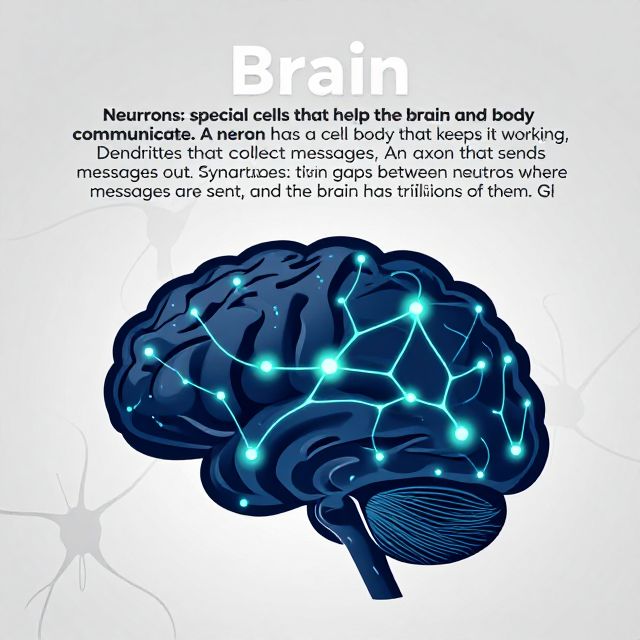
2. The Central Nervous System (CNS) 🧠

The CNS is the command center, consisting of the brain and the spinal cord. While the spinal cord is a single, continuous structure, the brain is organized into four principal regions: the Cerebrum, Diencephalon, Brainstem, and Cerebellum.

2.1. The Cerebrum

The cerebrum is the largest part of the brain, responsible for higher-level functions.

  • Cerebral Cortex: The outer layer of gray matter, involved in consciousness, thought, memory, and voluntary movement.
  • Subcortical Nuclei: Deep structures within the cerebrum.
    • 📚 Basal Nuclei: Crucial for cognitive processing and the planning of movements. They compare cortical activity with overall nervous system activity to help decide if a movement should occur.
    • 📚 Basal Forebrain: Important for learning and memory. Its nuclei produce acetylcholine, regulating cortical activity and enhancing attention to sensory stimuli.
    • 📚 Limbic Cortex: Involved in emotion, memory, and behavior.
    • 📚 Hippocampus and Amygdala: Integral to long-term memory formation and emotional responses.

2.1.1. Basal Nuclei Pathways & Motor Disorders ⚠️

The basal nuclei are interconnected with brainstem nuclei, forming a vital motor pathway. This pathway involves two processing routes:

  • Direct Pathway: Generally facilitates movement.
  • Indirect Pathway: Generally inhibits movement. All input to this system originates from the cortex and projects into the striatum. A delicate balance between these pathways is essential for smooth, controlled movement. Damage to any part can lead to severe motor problems:
  • Bradykinesia: Slow movement.
  • Akinesia: Absence of movement.
  • Hypertonia: Muscle rigidity.
  • Dyskinesia: Involuntary movements. Examples: Parkinson's disease (PD) and Huntington's disease (HD) are notable disorders associated with basal nuclei dysfunction.

2.2. The Diencephalon

Located between the cerebrum and the brainstem, the diencephalon includes:

  • 📚 Thalamus: The main relay station for sensory and motor information to the cerebral cortex.
    • Relay Nuclei: Receive sensory, motor, and limbic input and send information to the cortex.
    • Association Nuclei: Connect different cortical areas.
    • Intralaminar Nuclei: Connected to the basal ganglia and limbic system.
    • Reticular Nucleus: Helps conscious awareness and synchronizes thalamus and cortex activity.
  • 📚 Hypothalamus: A vital regulatory center that controls numerous homeostatic functions:
    • Sets body temperature.
    • Controls blood pressure.
    • Manages body fluid balance.
    • Oversees autonomic functions.
    • Regulates sexual behavior, hunger, and thirst.
  • Epithalamus: Contains the pineal gland, involved in circadian rhythms.
  • Subthalamus: Contains the subthalamic nucleus, part of the basal nuclei.

2.3. The Brainstem

The brainstem connects the cerebrum and cerebellum to the spinal cord. It comprises three main parts:

  • Midbrain: Coordinates visual, auditory, and other sensory information.
  • Pons: Connects to the cerebellum and, along with the medulla, controls the cardiovascular and respiratory systems.
  • Medulla Oblongata: Regulates vital autonomic functions like breathing, heart rate, and blood pressure.
  • Cranial Nerves: Many cranial nerves pass through the brainstem, governing head and neck functions.
  • Pathways: Ascending (sensory) and descending (motor) pathways traverse this region, linking the brain and spinal cord.

2.4. The Cerebellum

Often called the "little brain," the cerebellum is critical for motor control and coordination.

  • Functions:
    • Coordinates movement and balance.
    • Predicts movement results and adjusts ongoing movements.
    • Plays a role in language and cognition.
    • Compares information from the cerebrum with sensory feedback from the body to fine-tune and smooth learned movements.
  • Sensory Input: Receives extensive sensory data from muscles, tendons, joints, skin, visual system, and vestibular system.
  • Cerebellar Damage: Does NOT cause paralysis but results in ataxia (poor coordination), characterized by:
    • Staggering gait.
    • Slurred speech.
    • Swallowing difficulties.
    • Eye movement problems.

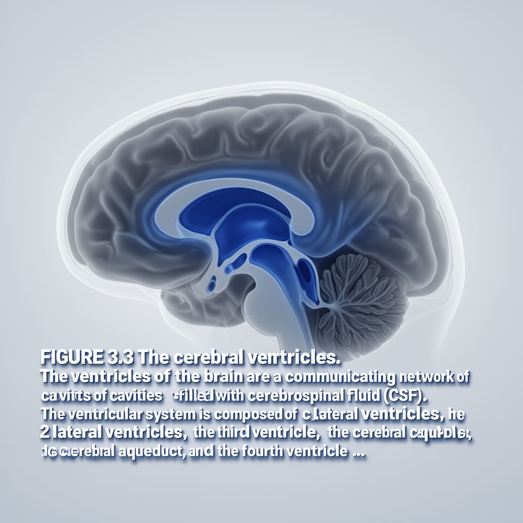
2.5. The Spinal Cord

The spinal cord is a long, slender bundle of nervous tissue extending from the brainstem.

  • Functional Regions:
    • Posterior Regions: Primarily responsible for sensory functions.
    • Anterior Regions: Primarily associated with motor functions.

3. The Peripheral Nervous System (PNS) 🌐

The PNS extends beyond the CNS, encompassing nerves and ganglia that transmit information throughout the body.

3.1. Ganglia

📚 Ganglia are clusters of nerve cell bodies located outside the CNS.

  • Sensory Ganglia:
    • Dorsal Root Ganglion: Most common type, associated with spinal nerves.
    • Cranial Nerve Ganglion: Associated with cranial nerves.
  • Autonomic Ganglia: Part of the autonomic nervous system.
    • Sympathetic Division: Includes sympathetic chain ganglia, paravertebral ganglia, and prevertebral ganglia. These regulate organs in the head, neck, thorax, abdomen, and pelvis.
    • Parasympathetic Division: Primarily uses terminal ganglia, receiving input from cranial or sacral nerves.

3.2. Nerves

📚 Nerves are bundles of nerve fibers (axons) that transmit electrical impulses. They contain nervous tissue, connective tissue, and blood vessels. Nerves are classified based on several criteria:

  • Structure:
    • Myelinated: Covered in a myelin sheath, allowing faster impulse conduction.
    • Non-myelinated: Lacking a myelin sheath, resulting in slower conduction.
  • Distribution:
    • Somatic: Innervate skeletal muscles and skin (voluntary control).
    • Visceral (Autonomic): Innervate internal organs, smooth muscle, and glands (involuntary control).
  • Origin:
    • Cranial Nerves: Attached directly to the brain.
    • Spinal Nerves: Connected to the spinal cord.
  • Function:
    • Sensory (Afferent): Carry sensory information to the CNS.
    • Motor (Efferent): Carry motor commands from the CNS.
  • Neurotransmitter Type:
    • Adrenergic: Release norepinephrine.
    • Cholinergic: Release acetylcholine.
  • Diameter and Conduction: Nerve fibers are also classified by their diameter and the speed at which they conduct impulses (e.g., A, B, C fibers).

3.2.1. Cranial Nerves

  • Attached to the brain, primarily control head and neck functions.
  • 💡 Insight: One notable cranial nerve, the Vagus nerve (Cranial Nerve X), extends to thoracic and abdominal organs, forming a significant part of the parasympathetic system.

3.2.2. Spinal Nerves

  • Connected to the spinal cord and extend from the vertebral column to the periphery.
  • Nerve Plexuses: Axons from different spinal nerves often join together to form systemic nerves at specific locations called plexuses (e.g., cervical, brachial, lumbar, sacral plexuses).
  • Other spinal nerves directly correspond to their vertebral levels.

4. Conclusion ✅

The nervous system, with its intricate Central and Peripheral divisions, orchestrates every aspect of our being. The CNS, comprising the brain and spinal cord, serves as the command center, integrating complex information and initiating responses through specialized regions like the cerebrum, diencephalon, brainstem, and cerebellum. Each area contributes uniquely to cognitive processing, motor control, sensory integration, and vital physiological regulation. The PNS, through its vast network of nerves and ganglia, acts as the essential communication link, relaying sensory input to the CNS and transmitting motor commands to the body's effectors. The precise interplay and anatomical integrity of these divisions are fundamental for coordinated bodily function and overall health.

Kendi çalışma materyalini oluştur

PDF, YouTube videosu veya herhangi bir konuyu dakikalar içinde podcast, özet, flash kart ve quiz'e dönüştür. 1.000.000+ kullanıcı tercih ediyor.

Sıradaki Konular

Tümünü keşfet
The Nervous and Endocrine Systems: Body's Control Centers

The Nervous and Endocrine Systems: Body's Control Centers

Explore the intricate workings of the nervous and endocrine systems, their structures, functions, and how they maintain the body's homeostasis.

Özet 25 15 Görsel
The Neural Bases of Behaviour: An Introduction

The Neural Bases of Behaviour: An Introduction

An introductory guide to how the nervous system and brain support human behaviour, covering the CNS, PNS, and the role of neurons.

3 dk 22 10
The Nervous System: Structure, Tissue, and Communication

The Nervous System: Structure, Tissue, and Communication

An academic overview of the nervous system's anatomical and functional divisions, its cellular components, the role of myelination, and the electrical and chemical mechanisms of neuronal communication.

7 dk Özet 25 15
The Developing Brain: Structure, Function, and Growth

The Developing Brain: Structure, Function, and Growth

Explore the intricate structure of the brain, its cellular components, major regions, and the complex processes of brain development and physical growth from conception to adulthood.

Özet 25 15
Nervous System: Structure, Function, and Signaling

Nervous System: Structure, Function, and Signaling

An academic overview of the nervous system's anatomical and functional divisions, cellular components, electrical signaling mechanisms, and inter-neuronal communication.

5 dk Özet 25 15
Human Brain Anatomy: Ventricles, Terminology, and Structures

Human Brain Anatomy: Ventricles, Terminology, and Structures

Explore the intricate anatomy of the human brain, covering the ventricular system, essential directional terms, embryonic development, and the major divisions and structures of the adult brain.

25 15
Neurolinguistics: Language, Brain, and Aphasia

Neurolinguistics: Language, Brain, and Aphasia

This audio summary explores neurolinguistics, its interdisciplinary nature, the structure and functions of the brain's language areas, and various types of aphasia resulting from brain damage.

6 dk Özet 25 15
Sensory and Motor Systems: An Academic Overview

Sensory and Motor Systems: An Academic Overview

This audio summary provides a formal academic overview of the human sensory and motor systems, detailing sensory perception, receptor types, neural pathways, motor control, and reflexes.

7 dk Özet 25 15