Sensory and Motor Systems: An Academic Overview - kapak
Bilim#sensory systems#motor systems#neurophysiology#proprioception

Sensory and Motor Systems: An Academic Overview

This audio summary provides a formal academic overview of the human sensory and motor systems, detailing sensory perception, receptor types, neural pathways, motor control, and reflexes.

aleyblgnMarch 29, 2026 ~24 dk toplam
01

Sesli Özet

8 dakika

Konuyu otobüste, koşarken, yolda dinleyerek öğren.

Sesli Özet

Sensory and Motor Systems: An Academic Overview

0:007:30
02

Flash Kartlar

25 kart

Karta tıklayarak çevir. ← → ile gez, ⎵ ile çevir.

1 / 25
Tüm kartları metin olarak gör
  1. 1. What is the primary role of the human body's sensory and motor systems?

    The sensory and motor systems are fundamental for interacting with the environment and executing movements. The sensory system detects environmental changes and converts stimuli into electrical signals for the CNS, while the motor system facilitates voluntary movement and maintains posture. They are interconnected and crucial for overall physiological function.

  2. 2. How are sensory receptors classified based on the stimuli they detect?

    Sensory receptors are classified into several types based on the specific stimuli they detect. These include mechanoreceptors for touch, pressure, and vibration; thermoreceptors for temperature; chemoreceptors for chemical stimuli; and nociceptors for pain sensation. This classification helps in understanding how different types of environmental information are initially processed.

  3. 3. What is somatosensation and what modalities does it encompass?

    Somatosensation refers to a broad category of senses originating from the skin, muscles, joints, and connective tissues. It encompasses modalities such as touch, pressure, vibration, temperature, pain, and proprioception. This system provides the brain with detailed information about the body's physical state and its interaction with the external world.

  4. 4. Define proprioception and provide an example of its function.

    Proprioception is the body's capacity to perceive the position of body parts, joint movement, and muscle tension. It allows individuals to know where their limbs are in space without looking. An example is the ability to touch one's nose with closed eyes, demonstrating awareness of arm and hand position.

  5. 5. Name three key proprioceptors and briefly describe their roles.

    Key proprioceptors include muscle spindles, Golgi tendon organs, and joint receptors. Muscle spindles detect changes in muscle length, providing information about muscle stretch. Golgi tendon organs sense muscle tension, protecting muscles from excessive force. Joint receptors monitor joint position and movement, contributing to overall body awareness.

  6. 6. What are some potential causes and symptoms of a loss of proprioception?

    A loss of proprioception can be caused by conditions such as diabetic neuropathy, vitamin B12 deficiency, spinal cord injury, and various neurological disorders. Symptoms often include poor balance, difficulty coordinating movements, and an increased risk of falls. This impairment significantly affects an individual's ability to navigate their environment safely.

  7. 7. How does thermoreception differentiate between cold and warm stimuli, and which fibers transmit this information?

    Thermoreception involves specialized cold and warm receptors, with cold receptors being more numerous in the skin. These receptors detect changes in temperature. The information gathered by these receptors is then transmitted to the central nervous system via Aδ and C fibers, allowing the brain to perceive temperature sensations.

  8. 8. Explain nociception, including the types of stimuli it detects and the fibers involved in transmitting pain signals.

    Nociception is the sensation of pain, mediated by nociceptors that detect actual or potential tissue damage. These stimuli can be mechanical, chemical, or thermal. Pain signals are transmitted through two types of fibers: Aδ fibers for fast, sharp pain, and C fibers for slow, dull, aching pain.

  9. 9. Name three organs that notably do not contain pain receptors.

    Certain internal organs, including brain tissue, liver tissue, and lung tissue, do not contain pain receptors. This means that direct injury to these specific tissues may not cause pain. Instead, pain associated with these organs often arises when surrounding structures, which do have nociceptors, are affected.

  10. 10. What is the primary function of the dorsal column pathway in sensory information transmission?

    The dorsal column pathway is a major ascending sensory pathway responsible for transmitting specific types of sensory information to the central nervous system. It primarily carries signals related to fine touch, proprioception (body position awareness), and vibration. This pathway is crucial for detailed tactile discrimination and spatial awareness.

  11. 11. What types of sensory information are transmitted via the spinothalamic tract?

    The spinothalamic tract is another vital ascending sensory pathway. Its primary function is to transmit signals related to pain and temperature. This pathway is essential for the body's protective responses to harmful stimuli and for maintaining thermal homeostasis.

  12. 12. Describe the general route sensory information takes from a receptor to the sensory cortex.

    The general route for sensory information involves a sequence of structures. It begins with the activation of a sensory receptor, which then transmits signals to the spinal cord. From there, the signals ascend to the thalamus, which acts as a relay station, before finally reaching the sensory cortex in the brain for conscious perception and processing.

  13. 13. What are the three stages of cortical processing for sensory information?

    Cortical processing of sensory information occurs in three distinct stages. First, the primary sensory cortex receives initial input, detecting stimulus location and intensity. Second, the sensory association area interprets the meaning of this information. Finally, the multimodal integration area combines sensory data with memory and experience for a comprehensive understanding.

  14. 14. Which brain regions are specialized for processing somatosensory, visual, and auditory input, respectively?

    Different brain regions are specialized for processing specific types of sensory input. The parietal lobe is primarily responsible for handling somatosensory input, such as touch and proprioception. The occipital lobe is dedicated to processing visual information, while the temporal lobe is specialized for auditory input.

  15. 15. Which lobe of the brain predominantly controls motor functions, and what are its three critical areas?

    Motor functions are predominantly controlled by the frontal lobe of the brain. Within the frontal lobe, three critical areas are involved: the primary motor cortex, the premotor cortex, and the supplementary motor area. These areas work together to plan, initiate, and execute voluntary movements.

  16. 16. What is the specific role of the primary motor cortex in movement?

    The primary motor cortex plays a crucial role in the initiation of voluntary movement. It generates the neural impulses that control the execution of movements. This area is directly involved in sending signals down to the spinal cord to activate specific muscles, allowing for conscious control over our actions.

  17. 17. Differentiate between upper motor neurons and lower motor neurons in the motor system hierarchy.

    The motor system operates with a hierarchical structure involving upper and lower motor neurons. Upper motor neurons are located in the brain (e.g., motor cortex) and transmit signals down to the spinal cord. Lower motor neurons are situated in the spinal cord or brainstem and directly stimulate skeletal muscles, acting as the final common pathway for motor commands.

  18. 18. What is the primary responsibility of the pyramidal system, and what tracts does it include?

    The pyramidal system is primarily responsible for voluntary movement, particularly fine and skilled movements. It includes two main tracts: the corticospinal tract and the corticobulbar tract. The corticospinal tract controls movements of the limbs and trunk, while the corticobulbar tract controls movements of the head and face.

  19. 19. Describe the specific function of the corticospinal tract.

    The corticospinal tract is a key component of the pyramidal system, originating in the motor cortex and projecting to the spinal cord. Its specific function is to control fine voluntary movements, especially skilled movements of the hands and fingers. This tract is essential for tasks requiring precision and dexterity.

  20. 20. What are the main functions of the extrapyramidal system, and what structures does it work with?

    The extrapyramidal system is responsible for regulating posture, balance, muscle tone, and automatic movements. It operates in conjunction with several subcortical structures, including the basal ganglia, cerebellum, and brainstem. This system ensures smooth, coordinated movements and maintains body stability.

  21. 21. Briefly explain the function of the tectospinal tract.

    The tectospinal tract is an extrapyramidal tract that originates in the superior colliculus of the midbrain. Its primary function is to mediate reflex movements of the head and neck. These movements are typically in response to visual stimuli, allowing for rapid orientation towards objects in the visual field.

  22. 22. What is the role of the reticulospinal tract in motor control?

    The reticulospinal tract, originating in the reticular formation of the brainstem, plays a significant role in controlling posture, regulating muscle tone, and influencing locomotion. It has both pontine and medullary components, which often have opposing effects on muscle tone, contributing to the complex regulation of motor activity.

  23. 23. How does the vestibulospinal tract contribute to motor function?

    The vestibulospinal tract is crucial for maintaining balance and posture. It comprises lateral and medial types. The lateral vestibulospinal tract primarily activates extensor muscles to counteract gravity and maintain upright posture, while the medial vestibulospinal tract helps coordinate head and eye movements.

  24. 24. What are the typical clinical manifestations of upper motor neuron lesions?

    Lesions to upper motor neurons, often seen in conditions like stroke or spinal cord injury, typically result in a characteristic set of symptoms. These include spastic paralysis, where muscles are stiff and resistant to movement, and increased reflexes (hyperreflexia). There may also be a positive Babinski sign.

  25. 25. What are the typical clinical manifestations of lower motor neuron lesions?

    Lower motor neuron lesions, which can occur due to peripheral nerve injury or diseases like poliomyelitis, present with different clinical signs. These typically include muscle weakness, muscle atrophy (wasting), and decreased or absent reflexes (hyporeflexia or areflexia). Fasciculations (muscle twitching) may also be observed.

03

Bilgini Test Et

15 soru

Çoktan seçmeli sorularla öğrendiklerini ölç. Cevap + açıklama.

Soru 1 / 15Skor: 0

Which type of sensory receptor is responsible for detecting changes in touch, pressure, and vibration?

04

Detaylı Özet

6 dk okuma

Tüm konuyu derinlemesine, başlık başlık.

📚 Sensory and Motor Systems: A Comprehensive Study Guide

Source Information: This study material has been compiled and organized from a lecture audio transcript and a copy-pasted text document provided by the user.


🧠 Introduction to Sensory and Motor Systems

The human body's ability to interact with its environment and execute movements relies fundamentally on its sensory and motor systems. These two interconnected systems work in harmony to allow us to perceive the world around us and respond to it. Sensory perception enables the detection of both external and internal environmental changes, converting various stimuli into electrical signals that are transmitted to the central nervous system (CNS). Concurrently, the motor system facilitates voluntary movement, maintains posture, and coordinates complex actions. This guide will delineate the components and functions of these vital systems, from receptor-level detection to cortical processing and motor execution.


👂 The Sensory System: Perceiving the World

The sensory system is responsible for gathering information from our environment and our own body.

1. Sensory Perception

📚 Sensory perception is the process by which the body detects changes in both the external and internal environments. ✅ Mechanism: Sensory receptors convert a stimulus (e.g., light, pressure, temperature) into an electrical signal, which is then transmitted to the central nervous system for processing. ✅ Classification: Senses can be broadly classified as either general (e.g., touch, pain) or specific (e.g., vision, hearing).

2. Sensory Receptors

Different types of stimuli are sensed by specialized receptor cells. These receptors can be classified based on the type of stimulus they detect:

  • Mechanoreceptors: Detect mechanical stimuli such as touch, pressure, and vibration.
  • Thermoreceptors: Detect changes in temperature (hot or cold).
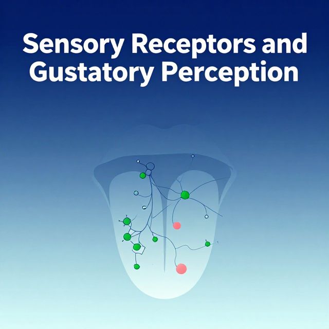
  • Chemoreceptors: Detect chemical stimuli, important for taste, smell, and internal chemical balances.
  • Nociceptors: Detect potentially damaging stimuli, leading to the sensation of pain.

3. Somatosensation

Somatosensation refers to the body senses, encompassing several sensory modalities originating from the skin, muscles, joints, and connective tissues. ✅ Modalities include: Touch, Pressure, Vibration, Temperature, Pain, and Proprioception.

Proprioception

📚 Proprioception is the body's ability to detect the position of body parts, the movement of joints, and muscle tension. It's our "sixth sense" of body awareness. 💡 Example: You can touch your nose with your eyes closed because of proprioception, knowing where your hand and nose are in space.

Proprioceptors

The main proprioceptors include:

  • Muscle Spindles: Detect changes in muscle length.
  • Golgi Tendon Organs: Detect muscle tension.
  • Joint Receptors: Detect joint position and movement. ✅ These receptors are essential for maintaining posture and coordination.
Loss of Proprioception

⚠️ A loss of proprioception can significantly impair movement and balance. It may occur in conditions such as:

  • Diabetic neuropathy
  • Vitamin B12 deficiency
  • Spinal cord injury
  • Various neurological disorders ✅ Symptoms may include poor balance, unstable walking, and an increased risk of falls.

4. Thermoreception

Thermoreceptors detect temperature changes.

  • Cold receptors: More numerous in the skin, responding to cooling.
  • Warm receptors: Less numerous, responding to warming. ✅ Temperature information is transmitted to the CNS via Aδ fibers (faster) and C fibers (slower).

5. Nociception (Pain Sensation)

Nociceptors detect actual or potential tissue damage.

  • Types of stimuli: Mechanical (e.g., a cut), Chemical (e.g., acid), Thermal (e.g., extreme heat/cold).
  • Pain signals travel through:
    • Aδ fibers: Transmit fast, sharp pain.
    • C fibers: Transmit slow, dull, aching pain. ⚠️ Important Note: Some organs, such as brain tissue, liver tissue, and lung tissue, do not contain pain receptors themselves. Pain from these areas occurs when surrounding structures (e.g., meninges around the brain, capsule around the liver) are affected.

6. Sensory Pathways

Sensory information travels through ascending pathways to the brain. 1️⃣ General Route: Receptor → Spinal cord → Thalamus → Sensory cortex. 2️⃣ Two Major Pathways:

  • Dorsal Column Pathway: Carries fine touch, proprioception, and vibration sensations.
  • Spinothalamic Tract: Carries pain and temperature sensations.

7. Cortical Processing of Sensory Information

Sensory information is processed in the cerebral cortex in three stages:

  1. Primary Sensory Cortex: Receives initial sensory input, detecting stimulus location and intensity.
  2. Sensory Association Area: Interprets the meaning of the sensory information (e.g., recognizing an object by touch).
  3. Multimodal Integration Area: Combines sensory information with memory and experience to form a complete perception. ✅ Brain Regions:
  • Parietal lobe: Primarily responsible for somatosensory processing.
  • Occipital lobe: Processes visual information.
  • Temporal lobe: Processes auditory information.

🏃 The Motor System: Movement and Control

The motor system is responsible for generating and controlling movements, from simple reflexes to complex voluntary actions.

1. Motor Cortex

Motor functions are mainly controlled by the frontal lobe, involving three important areas:

  • Primary Motor Cortex: Initiates voluntary movements.
  • Premotor Cortex: Controls posture and prepares muscles for movement.
  • Supplementary Motor Area: Plans complex movements and coordinates learned movements (e.g., playing a musical instrument).

2. Motor Control: Upper and Lower Motor Neurons

The motor system controls voluntary movement through a hierarchical structure:

  • Upper Motor Neurons (UMN): Located in the brain (motor cortex and brainstem). They initiate and regulate the activity of lower motor neurons.
  • Lower Motor Neurons (LMN): Located in the spinal cord or brainstem. They directly stimulate skeletal muscles to contract.

3. Motor Pathways (Descending Pathways)

Motor pathways are divided into two main systems:

a. Pyramidal System

✅ Responsible for voluntary movement.

  • Corticospinal Tract:
    • Controls fine voluntary movements, especially skilled movements of the hands and fingers.
    • Originates in the motor cortex and projects directly to the spinal cord.
  • Corticobulbar Tract: Controls voluntary movements of the face, head, and neck.

b. Extrapyramidal System

✅ Responsible for regulating posture, balance, muscle tone, and automatic movements. It works in conjunction with the basal ganglia, cerebellum, and brainstem.

  • Tectospinal Tract:
    • Origin: Superior colliculus (midbrain).
    • Function: Mediates reflex movements of the head and neck in response to visual stimuli.
    • 💡 Example: Turning your head toward a sudden flash of light.
  • Reticulospinal Tract:
    • Origin: Reticular formation in the brainstem.
    • Functions: Controls posture, regulates muscle tone, and influences locomotion.
    • Pontine Reticulospinal Tract: Increases muscle tone, activates extensor muscles.
    • Medullary Reticulospinal Tract: Decreases muscle tone, inhibits extensor muscles.
  • Vestibulospinal Tract:
    • Origin: Vestibular nuclei in the brainstem.
    • Function: Maintains balance and posture.
    • Lateral Vestibulospinal Tract: Activates extensor muscles, maintaining an upright posture.
    • Medial Vestibulospinal Tract: Coordinates head and eye movements.
  • Rubrospinal Tract:
    • Origin: Red nucleus (midbrain).
    • Function: Assists flexor muscle activity and helps with limb movement. Less prominent in humans compared to other species.

4. UMN and LMN Lesions

Damage to motor neurons results in distinct clinical signs:

  • UMN Lesion:
    • Examples: Stroke, multiple sclerosis, spinal cord injury.
    • Symptoms: Spastic paralysis (increased muscle tone), increased reflexes, positive Babinski sign.
  • LMN Lesion:
    • Examples: Peripheral nerve injury, poliomyelitis, neuropathy.
    • Symptoms: Muscle weakness, muscle atrophy (wasting), decreased or absent reflexes, fasciculations (muscle twitching).

⚡ Reflexes: Automatic Responses

📚 A reflex is a rapid, involuntary, and automatic response to a specific stimulus. Reflexes are crucial for protection and maintaining homeostasis.

1. Reflex Pathway

1️⃣ Stimulus: An external or internal change. 2️⃣ Receptor: Detects the stimulus. 3️⃣ Afferent Neuron: Transmits the signal to the spinal cord. 4️⃣ Integration Center: In the spinal cord, processes the signal (may involve interneurons). 5️⃣ Efferent Neuron: Transmits the signal from the spinal cord to the effector. 6️⃣ Motor Response: The effector (muscle or gland) responds. 💡 Example: The knee-jerk reflex, where tapping the patellar tendon causes the quadriceps muscle to contract.

2. Types of Reflexes

  • Stretch Reflex: Maintains muscle length and posture by causing a muscle to contract when it is stretched.
  • Golgi Tendon Reflex: Protects muscles from excessive tension by causing the muscle to relax when tension becomes too high.
  • Withdrawal Reflex: Removes a body part from a painful stimulus (e.g., quickly pulling your hand away from a hot stove).

✅ Conclusion

The sensory and motor systems are intricately linked, forming the foundation for an organism's interaction with its environment. Sensory perception, facilitated by diverse receptors and ascending pathways, allows for the detection and interpretation of various stimuli. Motor control, orchestrated by the motor cortex and descending pathways, enables precise and coordinated movements, alongside the maintenance of posture and balance. Reflexes provide rapid, automatic responses crucial for protection and basic motor function. Understanding these systems is fundamental to comprehending human physiology and neurological function.

Kendi çalışma materyalini oluştur

PDF, YouTube videosu veya herhangi bir konuyu dakikalar içinde podcast, özet, flash kart ve quiz'e dönüştür. 1.000.000+ kullanıcı tercih ediyor.

Sıradaki Konular

Tümünü keşfet
The Nervous System: Structure, Tissue, and Communication

The Nervous System: Structure, Tissue, and Communication

An academic overview of the nervous system's anatomical and functional divisions, its cellular components, the role of myelination, and the electrical and chemical mechanisms of neuronal communication.

7 dk Özet 25 15
The Nervous and Endocrine Systems: Body's Control Centers

The Nervous and Endocrine Systems: Body's Control Centers

Explore the intricate workings of the nervous and endocrine systems, their structures, functions, and how they maintain the body's homeostasis.

Özet 25 15 Görsel
Anatomy of the Nervous System

Anatomy of the Nervous System

An academic overview of the nervous system, detailing the Central Nervous System (CNS) and Peripheral Nervous System (PNS), their components, functions, and associated disorders.

8 dk Özet 25 15
Nervous System: Structure, Function, and Signaling

Nervous System: Structure, Function, and Signaling

An academic overview of the nervous system's anatomical and functional divisions, cellular components, electrical signaling mechanisms, and inter-neuronal communication.

5 dk Özet 25 15
Neuron Types and Nervous System Structures

Neuron Types and Nervous System Structures

This summary explores the diverse morphology and classification of neurons, their fundamental components, synaptic connections, and the crucial role of glial cells and peripheral nervous system structures.

6 dk Özet 25 15
Sensory Receptors and Gustatory Perception

Sensory Receptors and Gustatory Perception

This summary explores the structure, function, and mechanisms of various sensory receptors, including Merkel cells and different nerve endings, alongside the anatomy and molecular processes of taste buds.

7 dk Özet 25 15
Cerebellar Afferent and Efferent Pathways

Cerebellar Afferent and Efferent Pathways

Explore the intricate network of cerebellar afferent and efferent pathways, understanding how the cerebellum receives sensory input and modulates motor control.

6 dk Özet 25 15
Understanding the Borehole Environment

Understanding the Borehole Environment

This summary provides an academic overview of the borehole environment, detailing its characteristics, influencing factors, and significance in subsurface investigations and resource extraction.

6 dk Özet 15