📚 Cytology: The Study of the Cell and Its Division
Source Information: This study material is compiled from a lecture audio transcript and copy-pasted text (slides) provided by the user.
🎯 Introduction to Cytology
Cytology is the scientific study of cells, forming a fundamental pillar of modern biology. While Gregor Mendel's groundbreaking work on heredity was initially overlooked, the rise of cytology in the early 1900s provided the crucial cellular framework for understanding inheritance, particularly through the intricate processes of cell division. This period marked a significant shift towards comprehending how genetic material is accurately passed from one cellular generation to the next.
🦠 Bacterial Cell Division: Binary Fission
Bacteria, as prokaryotic organisms, exhibit a simpler yet highly efficient method of reproduction called binary fission.
Bacterial Genome
✅ Most bacteria possess a single, closed, circular chromosome that encodes nearly all essential functions for survival and reproduction. ✅ Many bacteria can also carry a variable number of smaller, circular DNA molecules called plasmids. These plasmids often confer specific advantages, such as antibiotic resistance or virulence factors, which are crucial in particular environments.
Process of Binary Fission
- Initiation: Binary fission begins at a specific site on the circular chromosome known as the Origin of Replication (Ori-C).
- Replication: DNA replication proceeds bidirectionally from Ori-C, moving in both directions around the circular DNA molecule.
- Termination: Replication continues until the two replication forks meet at a designated Termination Site (Ter-C), resulting in two complete and identical circular chromosomes.
- Cell Division: The division of the bacterial cell occurs at its midpoint, mediated by FtsZ proteins. These proteins form a ring-like structure, the Z-ring, precisely at the cell's midpoint (septum).
The Divisome Complex
📚 The Divisome Complex is a multi-protein assembly responsible for orchestrating bacterial cell division.
- FtsZ Ring: Acts as a central scaffold, recruiting other essential proteins.
- Recruited Proteins: Drive peptidoglycan synthesis (for the new cell wall), facilitate membrane constriction, and form the septum.
- Outcome: The coordinated action of these proteins ensures accurate and efficient division, producing two genetically identical daughter cells.
🧬 Eukaryotic Genome Organization
Eukaryotic cells exhibit a far more complex organization of genetic material compared to prokaryotes.
Chromosomes and Genome Size
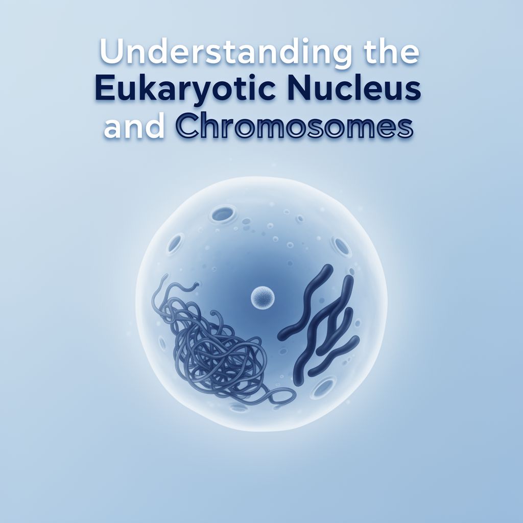
✅ In eukaryotes, DNA is intricately packed into highly organized structures called chromosomes. This packaging is vital for fitting vast amounts of genetic information into the nucleus and for accurate segregation during cell division. 💡 Genome Size vs. Complexity: Genome size is only loosely correlated with biological complexity. For example, Homo sapiens has a 3.3 Gbp genome, while Drosophila melanogaster has 122.6 Mbp. Some plants or amphibians can have much larger genomes without greater complexity, suggesting that factors like gene regulation play a significant role.
Mechanisms of Genome Expansion
Genomes are dynamic and can expand through various mechanisms:
- Polyploidization: An increase in the number of complete sets of chromosomes.
- Horizontal Gene Transfer (HGT): Transfer of genetic material between organisms not through parent-to-offspring inheritance (more common in bacteria but occurs in eukaryotes).
- DNA Duplications and Gene Family Expansion: Creates extra gene copies that can diverge and acquire new functions, contributing to genetic innovation.
Chromosome Structure and Terminology
- Appearance: Chromosomes appear as condensed, X-shaped structures during metaphase of cell division. However, during interphase (when the cell performs normal functions), DNA is much less condensed, existing as chromatin to allow gene expression and replication.
- Homologous Chromosomes: A typical human cell is diploid, possessing two copies of each chromosome (one from each parent). These pairs are called homologous chromosomes, carrying genes for the same traits at the same positions.
- Locus: The precise location of a specific gene on a chromosome. Gene loci are consistent within a species, allowing for the construction of genetic maps.
- Homology: Biological features descended from a feature present in a common ancestor.
- Orthologs: Genes in different species that evolved from a common ancestral gene by a speciation event (e.g., human and mouse hemoglobin genes).
- Paralogs: Genes within the same species that arose from a gene duplication event, often diverging to acquire new, related functions (e.g., different globin genes in humans).
- Ploidy Level: The number of sets of chromosomes in an organism.
- Humans are diploid (2n), with 46 chromosomes (23 pairs).
- Human gametes are haploid (n), with 23 different chromosomes.
- Karyotype: The complete set of chromosomes of an organism, organized and displayed. The human karyotype consists of 23 pairs of chromosomes (22 autosomes, 1 sex chromosome pair).
Chromatin Structure and Dynamics
📚 Chromatin: A complex of DNA (40%) and proteins (60%, mainly histones). The fundamental repeating unit is the nucleosome, where DNA is wound around a core of eight histone proteins.
- Chromatin Modifications (Epigenetics): Heritable changes in gene expression without altering the DNA sequence. Modifications (e.g., histone acetylation, DNA methylation) act as molecular switches, determining if a gene is "on" or "off" by influencing DNA accessibility.
- Euchromatin: Loosely packed, transcriptionally active regions (appears lighter with Giemsa staining).
- Heterochromatin: Tightly packed, generally transcriptionally inactive or silenced regions (appears darker with Giemsa staining).
- Nuclear Architecture:
- Chromosome Territories: Chromosomes occupy distinct regions within the nucleus.
- Compartments: Within territories, chromatin is organized into larger interacting regions.
- Topologically Associated Domains (TADs): Fundamental units of genome organization within compartments, characterized by high internal DNA-DNA interactions.
- TAD Anchoring: Anchored by the DNA-binding protein CTCF (insulator) and the cohesin complex (forms/stabilizes chromatin loops). This loop organization can bring distant regulatory elements together, affecting gene expression.
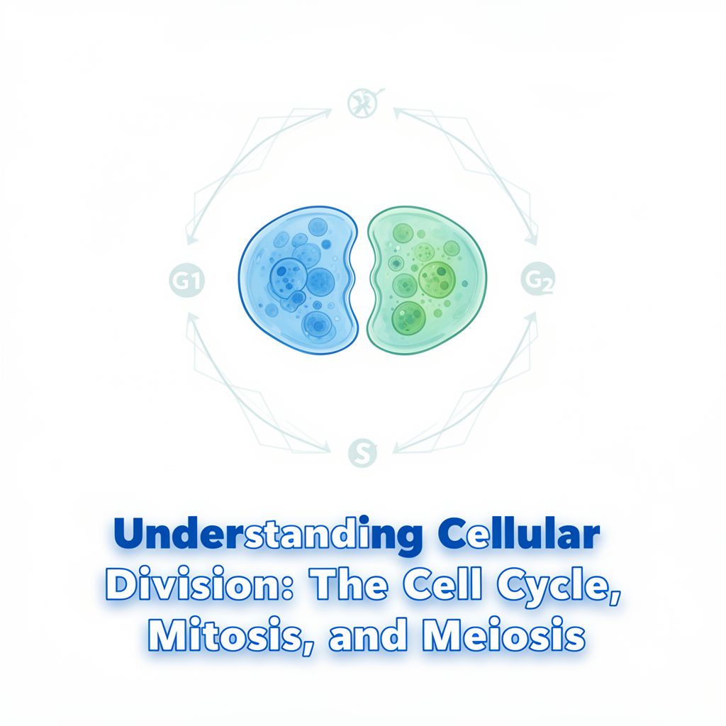
🔄 Eukaryotic Cell Cycle and Mitosis
The eukaryotic cell cycle is a precisely regulated sequence of events for growth, repair, and reproduction, involving genome duplication, accurate segregation, and cellular content division.
Phases of the Cell Cycle
The cell cycle consists of five main phases: G1, S, G2, M, and Cytokinesis. The first three (G1, S, G2) constitute Interphase.
- Interphase: The period of cell growth and DNA replication.
- G1 Phase (Growth 1): Cell grows, synthesizes proteins, and performs normal metabolic functions.
- S Phase (Synthesis): DNA replication occurs, resulting in two identical sister chromatids joined at the centromere.
- G2 Phase (Growth 2): Cell continues to grow, synthesizes proteins and organelles needed for division, and checks for DNA replication errors.
- M Phase (Mitosis): The dramatic period of nuclear division, a dynamic and continuous process broken down into:
- Prophase
- Prometaphase
- Metaphase
- Anaphase
- Telophase
- Cytokinesis: The division of the cytoplasm, typically following mitosis.
- In animal cells, a constricting belt of actin filaments (with myosin) forms a contractile ring and creates a cleavage furrow, eventually pinching the cell into two daughter cells.
Key Events in Mitosis
- Chromosome Condensation: Chromosomes become highly condensed during prophase and metaphase, making them compact and easier to move without tangling.
- Sister Chromatids: After replication, the two identical DNA molecules (sister chromatids) are held together at the centromere by cohesin proteins until separation.
- Mitotic Spindles: Responsible for organizing and sorting chromosomes. Composed of three types of microtubules:
- Astral microtubules: Anchor the spindle to the cell periphery.
- Interpolar microtubules: Extend from opposite poles, pushing them apart.
- Kinetochore microtubules: Attach directly to the kinetochores.
- Separation of Sister Chromatids: This crucial event depends on kinetochore microtubules attaching to the kinetochores (protein complexes at the centromere of each chromatid). These microtubules pull the sister chromatids apart towards opposite poles during anaphase.
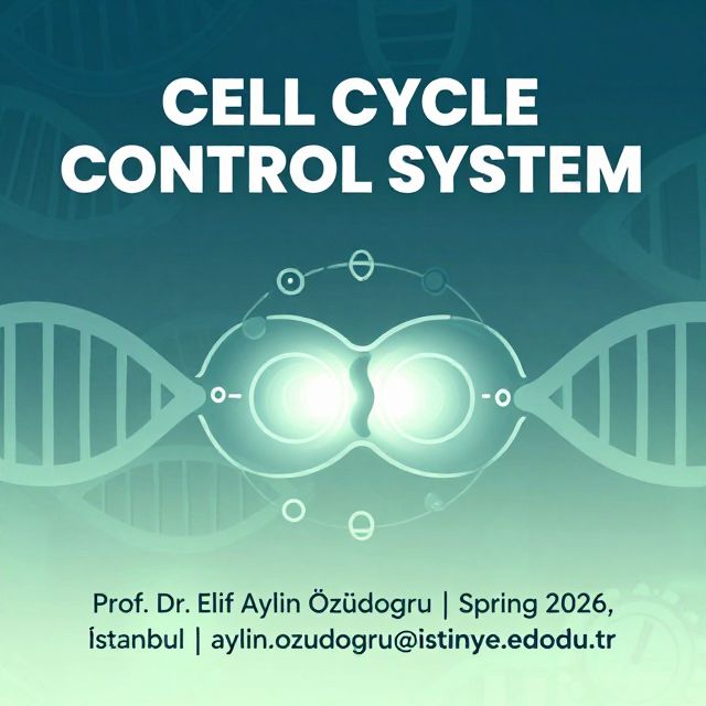
Cell Cycle Control and Checkpoints
⚠️ The entire cell cycle is governed by a sophisticated cell cycle control system, a network of regulatory proteins that monitors internal and external conditions.
- Irreversible Points: The cell cycle has two particularly irreversible points:
- Replication of genetic material (S phase).
- Separation of sister chromatids (Anaphase).
- Checkpoints: These processes are tightly controlled at specific checkpoints (e.g., G1, G2, M checkpoints). These surveillance mechanisms halt the cell cycle if errors are detected (e.g., unreplicated DNA, improperly attached chromosomes), ensuring genomic integrity and preventing mutations or chromosomal abnormalities.