Understanding Cellular Division: The Cell Cycle, Mitosis, and Meiosis - kapak
Bilim#cell division#cell cycle#mitosis#meiosis

Understanding Cellular Division: The Cell Cycle, Mitosis, and Meiosis

This podcast explores the intricate world of cellular division, detailing the cell cycle's phases and control, the process of mitosis for somatic cell reproduction, and meiosis for genetic diversity in gamete formation.

January 27, 2026 ~24 dk toplam
01

Sesli Özet

5 dakika

Konuyu otobüste, koşarken, yolda dinleyerek öğren.

Sesli Özet

Understanding Cellular Division: The Cell Cycle, Mitosis, and Meiosis

0:005:00
02

Flash Kartlar

25 kart

Karta tıklayarak çevir. ← → ile gez, ⎵ ile çevir.

1 / 25
Tüm kartları metin olarak gör
  1. 1. What is the fundamental purpose of cellular reproduction in living organisms?

    Cellular reproduction is fundamental to life, serving multiple critical purposes. It enables the growth of multicellular organisms from a single cell, allows for the repair and replacement of damaged or old tissues, and ensures the continuation of species through the production of new individuals. This process is essential for maintaining the integrity and functionality of living systems.

  2. 2. What crucial event must occur before a cell can divide, and what is its outcome?

    Before a cell can divide, it must undergo DNA duplication, a process that occurs during the S phase of interphase. This crucial event transforms single-chromatid chromosomes into double-chromatid chromosomes, meaning each chromosome now consists of two identical sister chromatids. The outcome is a precise doubling of the cell's genetic material, ensuring that each daughter cell receives a complete and identical set of chromosomes.

  3. 3. Name the two main stages of the cell cycle and their primary functions.

    The cell cycle comprises two main stages: interphase and mitosis (or M phase). Interphase is the longest phase, during which the cell grows, replicates its DNA, and prepares for division. Mitosis, or the M phase, is the actual division phase where the duplicated chromosomes are precisely separated into two new nuclei, followed by cytoplasmic division.

  4. 4. Describe the three distinct phases of interphase and the key activities occurring in each.

    Interphase consists of three distinct phases: G1, S, and G2. In the G1 phase (Gap 1), the cell grows and synthesizes proteins and organelles, preparing for DNA replication. The S phase (Synthesis) is when DNA replication occurs, doubling the genetic material. Finally, in the G2 phase (Gap 2), the cell continues to grow, synthesizes proteins necessary for mitosis, and prepares for cell division.

  5. 5. What is the G0 phase, and why do some cells enter it?

    The G0 phase is a quiescent or resting state that some cells enter, temporarily or permanently, when they are not actively dividing. Cells in G0 are metabolically active but have exited the cell cycle and are not preparing for DNA replication or division. This phase is common for highly differentiated cells like neurons or mature muscle cells, which typically do not divide.

  6. 6. What can be the consequence of deregulation at cell cycle restriction points?

    Deregulation at cell cycle restriction points can have severe consequences, primarily leading to uncontrolled cell growth and division. When these checkpoints fail to properly monitor and halt progression in the presence of errors, cells with damaged DNA or other abnormalities can continue to divide. This uncontrolled proliferation is a hallmark characteristic of malignant cells and cancer development.

  7. 7. What is the primary role of checkpoints in the cell cycle?

    The primary role of checkpoints in the cell cycle is to ensure the accurate and orderly progression through each phase. These checkpoints act as surveillance mechanisms, monitoring internal and external conditions, such as DNA integrity, chromosome alignment, and cell size. They prevent the cell from moving to the next phase until all previous steps are correctly completed, thereby maintaining genomic stability.

  8. 8. Identify three critical checkpoints that regulate the progression of the cell cycle.

    Three critical checkpoints regulate the progression of the cell cycle. The R or START checkpoint in G1 determines if the cell is ready to commit to division. The G2/M transition checkpoint ensures that DNA replication is complete and DNA is undamaged before entering mitosis. Finally, the metaphase-anaphase transition checkpoint verifies that all chromosomes are properly attached to the spindle fibers before sister chromatid separation.

  9. 9. Explain the function of cyclin-dependent kinases (cdks) in cell cycle regulation.

    Cyclin-dependent kinases (cdks) are a family of protein kinases that play a central role in regulating the cell cycle. They are enzymes that phosphorylate other proteins, thereby activating or inactivating them, to drive cell cycle progression. Cdks are only active when bound to specific regulatory proteins called cyclins, hence their name.

  10. 10. What are cyclins, and how do they interact with cdks to control the cell cycle?

    Cyclins are a family of proteins that fluctuate in concentration throughout the cell cycle, acting as regulatory subunits for cyclin-dependent kinases (cdks). They bind to and activate cdks, determining the substrate specificity and timing of cdk activity. The cyclical synthesis and degradation of cyclins ensure that cdk activity is precisely controlled, driving the cell through different phases of the cell cycle.

  11. 11. Provide an example of a cdk-cyclin complex and the cell cycle transition it regulates.

    An example of a cdk-cyclin complex is cdk2/3 associated with cyclin E, which is crucial for the G1-S transition. This complex phosphorylates target proteins that initiate DNA replication. Another example is cdk1 (also known as Cdc2) with cyclin B, which is essential for the G2/M transition, promoting entry into mitosis by phosphorylating proteins involved in nuclear envelope breakdown and spindle formation.

  12. 12. How do inhibitor proteins like p21 regulate the cell cycle?

    Inhibitor proteins, such as p21, regulate the cell cycle by inactivating cdk-cyclin complexes. They bind directly to these complexes, preventing the cdk from phosphorylating its target proteins. This inhibition can halt cell cycle progression, for instance, in response to DNA damage, allowing time for repair before the cell proceeds to the next phase.

  13. 13. Describe the role of the p53 protein in maintaining genomic integrity.

    The p53 protein is a critical tumor suppressor that acts as the "guardian of the genome." In response to DNA damage or other cellular stress, p53 becomes activated and can trigger cell cycle arrest, DNA repair, or apoptosis (programmed cell death). It achieves cell cycle arrest by activating the transcription of inhibitor genes, such as p21, which then blocks cdk activity.

  14. 14. What is the consequence of a mutated p53 protein on cell cycle progression?

    A mutated p53 protein loses its ability to detect DNA damage and activate cell cycle checkpoints or apoptosis. This failure means that cells with damaged DNA can continue to divide unchecked, leading to the accumulation of further mutations and genomic instability. Consequently, a mutated p53 is frequently found in human cancers, as it allows for permanent cdk activation and continuous cell cycle progression.

  15. 15. Explain the function of the Rb protein in cell proliferation control.

    The Retinoblastoma (Rb) protein acts as a crucial brake on cell proliferation, primarily by regulating the G1-S transition. In its active, unphosphorylated state, Rb binds to and inactivates transcription factors (like E2F) required for the expression of genes involved in DNA synthesis. This prevents the cell from entering the S phase until appropriate growth signals are received.

  16. 16. How does a mutation in the Rb protein contribute to uncontrolled cell division?

    A mutation in the Rb protein can lead to its inactivation, meaning it can no longer bind to and inhibit transcription factors like E2F. This loss of function removes the "brake" on cell proliferation, allowing cells to continuously progress through the G1-S checkpoint and divide without proper regulation. Such uncontrolled division is characteristic of various cancers, including retinoblastoma, from which the protein gets its name.

  17. 17. Why are p53 and Rb proteins referred to as anti-oncogenes?

    p53 and Rb proteins are referred to as anti-oncogenes, also known as tumor suppressor genes, because their normal function is to prevent uncontrolled cell growth and tumor formation. When these genes are functional, they regulate cell division, induce apoptosis in damaged cells, or repair DNA. Mutations that inactivate these proteins remove these protective mechanisms, thereby promoting cancer development.

  18. 18. Differentiate between direct and indirect cell division.

    Cell division occurs as either direct division, known as amitosis, or indirect division, which encompasses mitosis and meiosis. Direct division is a simpler process without a complex mitotic apparatus. Indirect division, on the other hand, involves precise and elaborate cytoplasmic and nuclear changes to ensure accurate distribution of genetic material.

  19. 19. What is amitosis, and in which organisms or tissues is it commonly observed?

    Amitosis is a form of direct cell division characterized by the absence of a mitotic apparatus and spindle formation. In this process, the nucleus and cytoplasm simply constrict and divide, often resulting in an unequal distribution of genetic material. Amitosis is commonly observed in unicellular organisms, rapidly dividing tissues, or in some specialized cells that do not require precise chromosome segregation.

  20. 20. Define mitosis and state its primary outcome for somatic cells.

    Mitosis is a type of indirect cell division that occurs in somatic cells, resulting in the production of two genetically identical diploid daughter cells from a single parent cell. Its primary purpose is to facilitate growth, tissue repair, and asexual reproduction in some organisms. This process ensures that each new cell receives a complete and accurate set of chromosomes.

  21. 21. List the five main phases of mitosis in chronological order.

    The five main phases of mitosis, in chronological order, are prophase, prometaphase, metaphase, anaphase, and telophase. These nuclear division phases are followed by cytokinesis, which is the division of the cytoplasm. Each phase involves distinct cellular events that contribute to the precise segregation of chromosomes.

  22. 22. Describe the key events that occur during prophase of mitosis.

    During prophase of mitosis, several key events occur. The chromatin condenses into visible, double-chromatid chromosomes, becoming shorter and thicker. The mitotic spindle begins to form from the centrosomes, which move to opposite poles of the cell. The nucleolus disappears, and the nuclear envelope starts to break down in later prophase (sometimes called prometaphase).

  23. 23. What is the defining event of metaphase in mitosis?

    The defining event of metaphase in mitosis is the alignment of all chromosomes at the metaphase plate, also known as the equatorial plate. Each chromosome, consisting of two sister chromatids, is positioned midway between the two spindle poles. This precise alignment ensures that when the sister chromatids separate, each daughter cell receives an identical set of genetic material.

  24. 24. Explain the crucial process that takes place during anaphase of mitosis.

    During anaphase of mitosis, the crucial process of sister chromatid separation takes place. The cohesin proteins holding sister chromatids together are cleaved, allowing the sister chromatids to rapidly pull apart. These now individual chromosomes are then moved by motor proteins along the spindle microtubules towards opposite poles of the cell, ensuring their equal distribution.

  25. 25. What happens during telophase and cytokinesis to complete cell division?

    Telophase marks the end of nuclear division, where the separated chromosomes arrive at the poles and begin to decondense. New nuclear envelopes form around the two sets of chromosomes, and the nucleoli reappear. Cytokinesis, the division of the cytoplasm, usually overlaps with telophase, forming a contractile ring of actin and myosin that pinches the cell into two distinct daughter cells.

03

Bilgini Test Et

15 soru

Çoktan seçmeli sorularla öğrendiklerini ölç. Cevap + açıklama.

Soru 1 / 15Skor: 0

Which phase of interphase is characterized by the synthesis of DNA, resulting in the doubling of genetic material?

04

Detaylı Özet

9 dk okuma

Tüm konuyu derinlemesine, başlık başlık.

This study material has been compiled from a copy-pasted text and an audio lecture transcript to provide a comprehensive overview of cellular division.


🧬 Cellular Division: The Cell Cycle, Mitosis, and Meiosis

1. Introduction to Cellular Division

Cellular division is a fundamental property of living organisms, essential for growth, repair, and reproduction. It is the process by which a parent cell divides to produce two or more daughter cells. Before division, a cell undergoes biochemical reproduction, doubling its components, including DNA, which transforms single-chromatid chromosomes into double-chromatid chromosomes. These sequential events form the cell cycle.
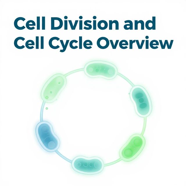
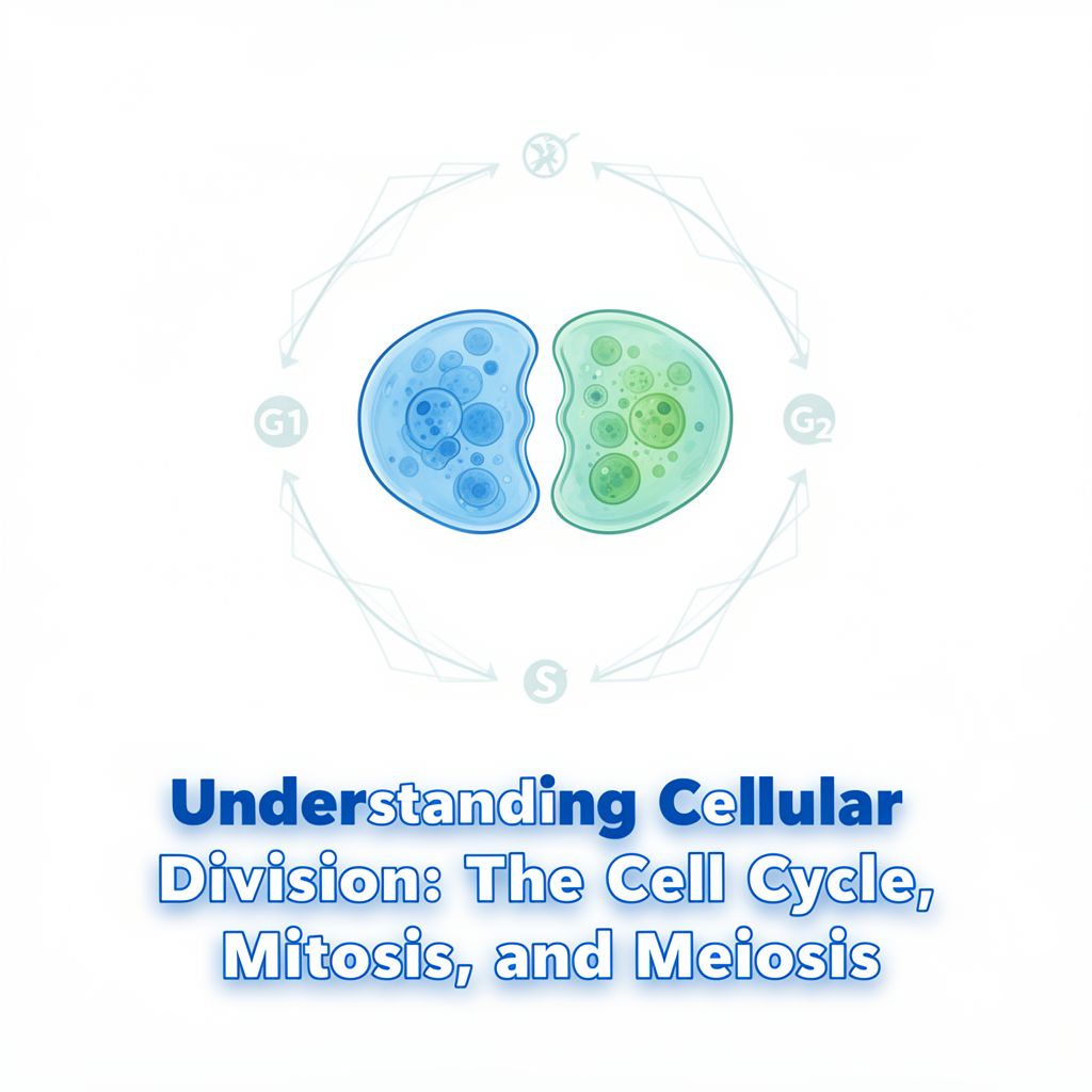
2. The Cell Cycle

📚 The cell cycle is defined as the period from the end of one cell division to the end of the subsequent division of the daughter cells. It consists of two main periods:

  • Interphase: The period between two consecutive cell divisions, where the cell grows and duplicates its DNA.
  • Mitosis (M phase): The period of nuclear and cytoplasmic division.

2.1. Phases of Interphase

Interphase is further divided into three sub-phases:

  • G1 Phase (Gap 1):
    • The cell grows and synthesizes proteins and organelles.
    • DNA amount remains constant (diploid, 2n single-chromatid chromosomes).
    • This phase is highly variable in length and can be the longest.
  • S Phase (Synthesis):
    • DNA replication occurs, doubling the genetic material.
    • Single-chromatid chromosomes become double-chromatid chromosomes.
    • The quantity of DNA doubles.
    • Typically lasts 7-8 hours.
  • G2 Phase (Gap 2):
    • The cell continues to grow and synthesizes proteins necessary for mitosis.
    • DNA amount remains constant (2n double-chromatid chromosomes).
    • Typically lasts 3-4 hours.

2.2. G0 Phase (Quiescence)

Some cells exit the cell cycle from G1 and enter a quiescent state called G0.

  • Cells in G0 can remain for extended periods (e.g., years).
  • Examples: Neurons, muscle cells, and some stomach cells.
  • 💡 Insight: Neurons, traditionally thought to be terminally differentiated, can sometimes be stimulated to divide under specific conditions, which could be relevant for neurodegenerative disease therapies.
  • Cells in G0 can re-enter the cell cycle if stimulated by mitogenic factors (e.g., growth factors, hormones).

2.3. Cell Classification by Division Capability

Cells can be categorized based on their cell cycle progression:

  1. Cells stopped in G1 (G0): Do not divide (e.g., mature neurons, muscle cells).
  2. Rapidly dividing cells: Continuously cycle (e.g., bone marrow, epidermis, intestinal epithelium, sperm germinal line). These tissues contain stem cells that replenish lost cells.
  3. Cells with low division capability: Divide only under special circumstances (e.g., endocrine cells, fibroblasts, liver cells, which can divide rapidly after injury to regenerate tissue).

3. Control of the Cell Cycle

The cell cycle is tightly regulated by a sophisticated system of checkpoints, ensuring proper completion of each phase before proceeding to the next. Deregulation of these controls can lead to malignant cell growth.

3.1. Key Checkpoints ✅

  • R or START Checkpoint (G1):
    • Determines if the cell is large enough and has sufficient growth factors to proceed.
    • Options: continue, pause, or enter G0.
  • G2/M Transition Checkpoint:
    • Ensures the cell has reached adequate size and DNA replication is complete and accurate.
  • Metaphase-Anaphase Transition Checkpoint:
    • Ensures all chromosomes are correctly attached to the mitotic spindle microtubules.

3.2. Molecular Regulators 🔬

Cell cycle progression is driven by the phosphorylation of proteins, primarily by enzymes called cyclin-dependent kinases (Cdks).

  • Cdks: These enzymes have a catalytic subunit and require an activator protein called a cyclin to be active.
  • Cyclins: Their concentration fluctuates cyclically throughout the cell cycle.
  • Cdk-Cyclin Complexes: Specific Cdk-cyclin pairs regulate different checkpoints:
    • G1-S Transition (START): Cdk2 or Cdk3 associates with Cyclin E.
    • G2/M Transition: Cdk1 associates with Cyclin B.
  • Inhibitor Proteins: Cdk-cyclin complexes can be inactivated by inhibitor proteins (e.g., p21, p16, p17 for G1).
  • p53 Protein (Tumor Suppressor):
    • Activated by DNA damage.
    • Activates the transcription of genes for Cdk inhibitor proteins (e.g., p21).
    • ⚠️ Warning: If p53 is mutated, it cannot activate these inhibitors, leading to permanent Cdk activation, uncontrolled cell cycle progression, and cancer (found in 50-60% of human cancers).
  • Rb Protein (Retinoblastoma Protein):
    • Acts as a brake on cell proliferation by binding to regulatory proteins, preventing gene expression for cell proliferation.
    • ⚠️ Warning: If Rb is mutated or absent, proliferation-stimulating proteins are continuously synthesized, leading to uncontrolled cell division, as seen in retinoblastoma.
  • Tumor Suppressor Genes: Both p53 and Rb are considered anti-oncogenes because they prevent uncontrolled cell growth.

4. Types of Cell Division

Cell division occurs in two main forms:

  1. Direct Division (Amitosis):
    • Lacks a mitotic apparatus.
    • The cell simply splits into two parts.
    • Common in unicellular organisms, or rapidly dividing/regenerating tissues (e.g., tumor cells).
  2. Indirect Division:
    • Involves synchronous cytoplasmic and nuclear changes.
    • Ensures equal distribution of genetic material to daughter cells.
    • Includes Mitosis and Meiosis.

5. Mitosis: Somatic Cell Division

Mitosis occurs in all somatic cells, producing two genetically identical daughter cells with the same chromosome number as the parent cell (e.g., a 2n cell produces two 2n cells).

5.1. Phases of Mitosis 📊

Mitosis is a continuous process, classically described in four phases, but often includes prometaphase and is followed by cytokinesis. Total duration is about 1 hour.

  1. Prophase (approx. 30 min):
    • Cytoplasm: Centrioles replicate, forming two centrosomes that move to opposite poles. The mitotic spindle (microtubules) begins to assemble between them. Asters form from centrosomes.
    • Nucleus: Nucleolus disassembles. Chromatin condenses into visible, double-chromatid chromosomes (each with two DNA molecules).
  2. Prometaphase:
    • Nuclear envelope disappears.
    • Mitotic apparatus fully develops.
    • Chromosomes undergo undulating movements, moving towards the metaphase plate.
  3. Metaphase (approx. 8 min):
    • Chromosomes align at the metaphase plate (equator of the cell), with their long axes perpendicular to the spindle axis.
    • At the end, sister chromatids separate at the centromere, becoming individual single-chromatid chromosomes.
  4. Anaphase (approx. 4 min):
    • Single-chromatid chromosomes move along the mitotic spindle towards opposite poles.
  5. Telophase (approx. 18 min):
    • Chromosomes arrive at the poles.
    • Nuclear envelopes reassemble around each set of chromosomes.
    • Nucleoli reappear.
    • Chromosomes decondense, returning to chromatin form.
    • Mitotic apparatus disassembles.
  6. Cytokinesis:
    • Cytoplasmic division, typically starting during anaphase or telophase.
    • A cleavage furrow forms on the plasma membrane, which deepens due to a contractile ring of actin and myosin, eventually pinching the cell into two daughter cells.

5.2. Molecular Mechanisms of Mitosis 💡

  • Centriole Replication: Occurs in S phase, completed in prophase, forming two centrosomes.
  • Mitotic Spindle Formation: Microtubules depolymerize from the cytoskeleton and reassemble into the spindle under centrosome control.
    • Astral microtubules: Extend from centrosomes to the plasma membrane.
    • Interpolar microtubules: Overlap at the spindle equator, pushing poles apart.
    • Kinetochore microtubules: Attach to kinetochores (specialized protein structures on centromeres) of chromosomes.
  • Chromosome Movement: Driven by molecular motor proteins (e.g., kinesins, dyneins) that "walk" along microtubules, consuming ATP.
    • Kinesin-related proteins (KRPs) move chromosomes along kinetochore microtubules.
    • Proteins at centrosomes pull kinetochore microtubules.
    • Shortening of kinetochore microtubules by depolymerization pulls chromosomes to poles.
    • Elongation of interpolar microtubules pushes centrosomes apart.
    • Cytosolic dyneins linked to the plasma membrane pull centrosomes towards the membrane.
  • Nuclear Envelope Disassembly/Reassembly: Phosphorylation of nuclear lamins (A, B, C) causes disassembly in prophase; dephosphorylation in telophase allows reassembly.
  • Cytokinesis: Contractile ring of actin and myosin forms a cleavage furrow, driven by myosin's motor activity.
  • Plasma Membrane Biogenesis: Intensified before division to provide sufficient membrane for two daughter cells.

5.3. Types of Mitosis

Based on the similarity between parent and daughter cells:

  1. Homotypic Mitosis: Daughter cells are identical to the mother cell.
  2. Heterotypic Mitosis (Differentiation Mitosis): Daughter cells are similar but more differentiated (mature) than the mother cell.
  3. Asymmetric Mitosis: One daughter cell is similar to the mother, the other is different.
  4. Dedifferentiation Mitosis: Daughter cells are younger/less differentiated than the mother cell (e.g., in lymphocytes).

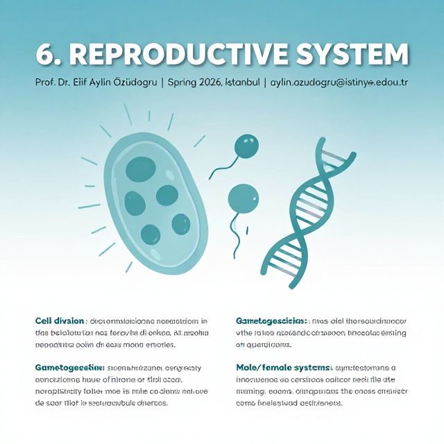
6. Meiosis: Gamete Formation and Genetic Diversity

Meiosis is a specialized type of cell division that reduces the chromosome number by half, producing four haploid daughter cells from a single diploid parent cell. It is crucial for sexual reproduction and genetic diversity.

6.1. Overview and Significance

  • Outcome: From one diploid (2n) mother cell, four haploid (n) daughter cells are produced.
  • Purpose: To form gametes (sperm and egg cells).
  • Genetic Restoration: Ensures that when two haploid gametes fuse during fertilization, the characteristic diploid chromosome number of the species is restored.
  • Genetic Diversity: Introduces genetic variation through recombination.

6.2. Meiosis I and Meiosis II

Meiosis consists of two consecutive divisions, Meiosis I and Meiosis II, without an intervening S phase (no DNA synthesis between the two divisions).

6.2.1. Meiosis I (Reductional Division)

  • Homologous chromosomes separate, reducing the chromosome number by half.
  • Prophase I (Longest and most complex):
    • Chromatin Condensation: Chromatin condenses into meiotic chromosomes.
    • Synapsis: Homologous chromosomes pair up precisely, forming bivalents (or tetrads, as they contain four chromatids). This pairing is facilitated by a synaptonemal complex.
    • Crossing-Over: Reciprocal exchange of genetic material between non-sister chromatids of homologous chromosomes. This shuffles genes, creating new combinations and increasing genetic diversity. The points of exchange are called chiasmata.
    • Duration: Can last for months or years in oocytes.
  • Prometaphase I: Nuclear envelope disappears, spindle forms.
  • Metaphase I: Bivalents align at the metaphase plate.
  • Anaphase I: Homologous chromosomes (each still composed of two chromatids) separate and move to opposite poles.
  • Telophase I: Chromosomes arrive at poles, nuclear envelopes may reform, and cytokinesis divides the cell into two haploid daughter cells.

6.2.2. Meiosis II (Equational Division)

  • Similar to mitosis, where sister chromatids separate.
  • The two haploid cells from Meiosis I divide again.
  • Prophase II: Chromatin condenses, double-chromatid chromosomes appear.
  • Prometaphase II: Nuclear envelope disappears, chromosomes move to the equator.
  • Metaphase II: Chromosomes align at the metaphase plate, and sister chromatids separate.
  • Anaphase II: Single-chromatid chromosomes migrate to opposite poles.
  • Telophase II: Nuclear envelopes reform, and cytokinesis results in four haploid daughter cells, each with single-chromatid chromosomes.

6.3. Meiosis in Humans 🚻

  • Oogenesis (Female Gamete Formation):
    • Primary oocytes begin Meiosis I in utero and arrest in Prophase I until puberty.
    • One primary oocyte completes Meiosis I each month, producing a large secondary oocyte and a small first polar body (asymmetric division).
    • The secondary oocyte arrests in Metaphase II and is released during ovulation. It completes Meiosis II only upon fertilization, forming an ovum and a second polar body.
    • Aging Effect: The long duration of Prophase I (up to 50 years) increases the risk of chromosomal anomalies with maternal age due to prolonged exposure to mutagenic factors.
  • Spermatogenesis (Male Gamete Formation):
    • Spermatogonia divide by mitosis at puberty, forming primary spermatocytes.
    • Primary spermatocytes undergo Meiosis I to form secondary spermatocytes.
    • Secondary spermatocytes undergo Meiosis II to form spermatids, which then differentiate into sperm cells.
    • This process is continuous from puberty into advanced age.

6.4. Genetic Diversity from Meiosis 🌍

  • Crossing-Over: Exchange of genetic material between homologous chromosomes creates recombinant chromatids.
  • Independent Assortment: Random alignment and separation of homologous chromosomes during Meiosis I.
  • Random Fertilization: Any sperm can fertilize any egg. These mechanisms ensure that each gamete is genetically unique, leading to the incredible diversity observed within species.

Kendi çalışma materyalini oluştur

PDF, YouTube videosu veya herhangi bir konuyu dakikalar içinde podcast, özet, flash kart ve quiz'e dönüştür. 1.000.000+ kullanıcı tercih ediyor.

Sıradaki Konular

Tümünü keşfet
Cell Division and Cell Cycle: Meiosis

Cell Division and Cell Cycle: Meiosis

An academic summary of cell division, focusing on sexual reproduction, the necessity of meiosis, its stages, and unique features like synapsis, homologous recombination, and reduction division.

6 dk Özet 25 15
Introduction to Basic Genetics

Introduction to Basic Genetics

Explore the fundamental principles of genetics, from cell theory and the nature of genetic material to heredity, cell division, and the diverse branches of genetic study.

Özet 25 15
The Cell Cycle Control System Explained

The Cell Cycle Control System Explained

Explore the intricate mechanisms governing cell division, from key proteins like cyclins and Cdks to the checkpoints ensuring proper replication and mitosis.

Özet 25 15
Cytology: The Study of the Cell and Its Division

Cytology: The Study of the Cell and Its Division

Explore the fundamental principles of cytology, from bacterial binary fission to the intricate eukaryotic cell cycle, chromosome organization, and genetic mechanisms.

17 dk Özet 25 15
Cell Division and Cell Cycle Overview

Cell Division and Cell Cycle Overview

An academic summary detailing prokaryotic and eukaryotic cell division, including binary fission, the eukaryotic cell cycle phases, and the stages of mitosis and cytokinesis.

6 dk Özet 25 15
Gene and Genome: Fundamental Concepts

Gene and Genome: Fundamental Concepts

This audio summary provides an academic overview of genes and genomes, detailing their definitions, structural components, functional aspects, and organizational differences between prokaryotic and eukaryotic systems.

6 dk Özet 25 15
Cellular Biology: Foundations of Life

Cellular Biology: Foundations of Life

Explore the fundamental principles of cellular biology, from its historical evolution and basic cell types to the molecular components and essential processes that define life.

7 dk Özet 25 15
The Reproductive System: Cell Division and Reproduction

The Reproductive System: Cell Division and Reproduction

Explore the fundamental processes of cell division and the diverse strategies of reproduction, including asexual and sexual methods, gametogenesis, fertilization, and the human reproductive systems.

Özet 25 15 Görsel