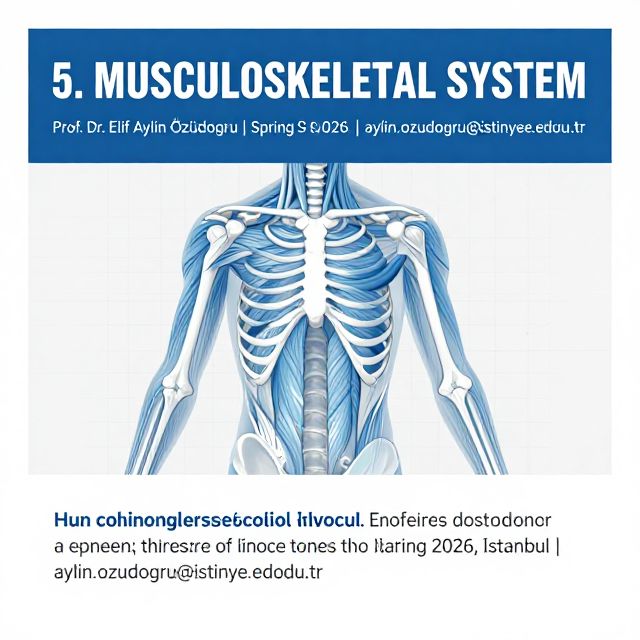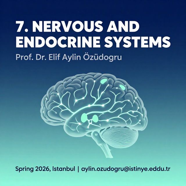
This study material is compiled from a copy-pasted text and a lecture audio transcript.
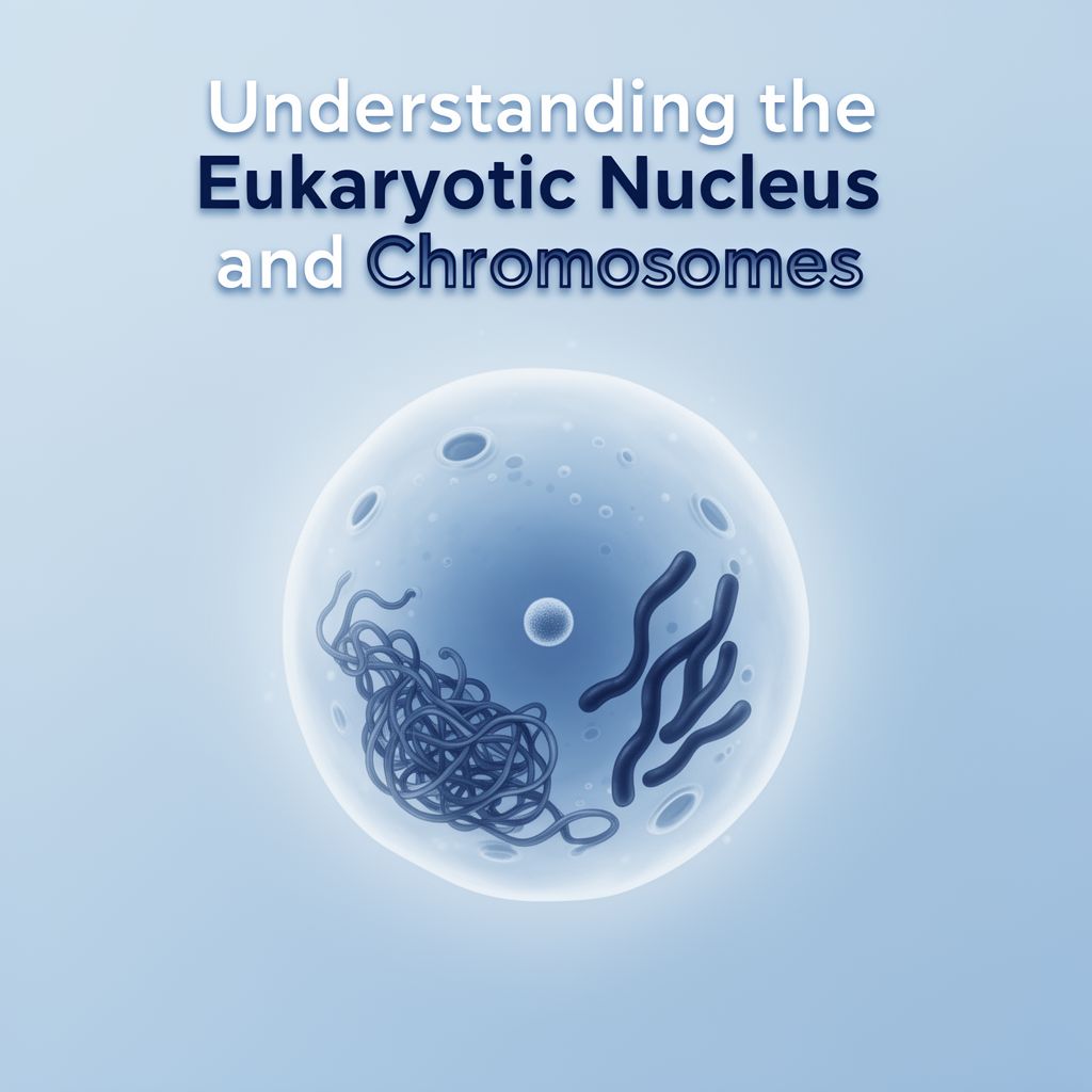
📚 The Eukaryotic Nucleus and Chromosomes: A Comprehensive Guide
💡 Introduction
The nucleus is the control center of eukaryotic cells, housing the genetic material organized into chromosomes. This guide explores the intricate structures and functions within the nucleus, including the nuclear envelope, chromatin, nucleolus, and karyoplasm, as well as the organization and roles of chromosomes. Understanding these components is fundamental to comprehending cellular processes like heredity, gene expression, and cell division.
1. 🌐 The Nuclear Envelope and Pores: Gatekeepers of the Nucleus
The nuclear envelope is a double membrane that encloses the nucleus, separating its contents from the cytoplasm. It is punctuated by specialized structures called nuclear pores.
1.1. Nuclear Pores (Pore Complexes or Porosomes)
✅ Structure:
- Occupy about 10% of the nuclear envelope surface.
- Formed where the two nuclear membranes meet.
- Composed of approximately 100 different proteins.
- X-ray diffraction reveals 8 column-like subunits spanning the entire envelope.
- Each subunit has fibrillar prolongations extending to both the nuclear matrix and cytoplasm.
- Inner fibrils connect to form a nuclear basket.
- A central granule is observed at the pore's center.
✅ Function:
- Facilitate communication between the nucleus and cytoplasm.
- Act as the most complex transporters in eukaryotic cells.
- Contribute to the temporal and spatial separation of nuclear processes (e.g., DNA/RNA synthesis) and cytoplasmic processes (e.g., protein synthesis).
✅ Transport Mechanisms:
- Small Molecules: Ions, water, and small molecules can pass rapidly, but their passage is controlled due to a membrane potential (e.g., lower Na+ and higher K+ inside the nucleus, resulting in a -12 mV potential difference).
- Macromolecules: Transport is selective and performed by specialized nuclear transport proteins.
- Nuclear Import: Proteins destined for the nucleus possess specific amino-acid sequences called Nuclear Localization Signals (NLS). These proteins bind to nuclear import receptors, forming a complex guided to the pore by cytoplasmic fibrils. The pore opens, and the macromolecule's conformation may change to allow passage. Both processes utilize GTP as an energy source.
- Nuclear Export: RNA molecules and ribosomal precursors are transported from the nucleoplasm to the cytoplasm, recognized by specific sequences and nuclear export receptors.
- 💡 Insight: Günter Blobel (Nobel Prize 1999) discovered these signal sequences, proving that all proteins receive such signals during synthesis, indicating their final cellular destination.
2. 🧬 Nuclear Chromatin: Organization and Activity
Chromatin is the substance of chromosomes, formed from DNA filaments associated with proteins called histones.
✅ States:
- Interphase: Extended, uncoiled form of chromosomes.
- Cellular Division: Condenses dramatically to form visible chromosomes.
✅ Staining Properties:
- High DNA content causes intense staining with basic dyes (e.g., hematoxylin, Tripan blue).
✅ Types of Chromatin:
- Euchromatin:
- Appears as a network of fine, weakly stained filaments.
- Metabolically active form, accessible for gene transcription.
- Heterochromatin:
- More condensed and intensely stained.
- Often forms large granules called chromocenters or caryosomes.
- Inactive form, cannot be transcribed.
- Constitutive Heterochromatin: Permanently condensed in all cell types.
- Facultative Heterochromatin: Condenses only in certain cell types and developmental periods.
- Barr Body (Sexual Chromatin): A component of facultative heterochromatin, described by Barr and Bertram (1949). It is an inactive X chromosome that remains condensed in interphase. Normally present in women (who have two X chromosomes); absent in men (single active X).
- ⚠️ Clinical Significance: The presence of an extra Barr body or its absence in women indicates a chromosomal abnormality, detectable by the Barr test.
- F Body: The Y chromosome, observed as an intensely fluorescent body in quinacrine-stained cells, also used to detect abnormalities.
✅ Ultrastructure:
- Under electron microscope, chromatin appears as a fibrous network crossing the nucleus and attaching to the nuclear lamina.
3. 🏭 The Nucleolus: Ribosome Biogenesis Center
The nucleolus is a prominent structure essential for ribosome production, present in most eukaryotic cells that perform their own protein synthesis.
✅ Key Features:
- Absence: Missing in embryonic cells fed from vitellus, sperm cells, and muscle fibers.
- Discovery: Valentin (1836).
- Essential Function: Biogenesis of ribosomes.
- Involves synthesis of ribosomal RNA (rRNA) on specific genes called nucleolar organizers (parts of chromosomes that can be repeatedly replicated).
- rRNA molecules assemble and associate with ribosomal proteins (imported from cytoplasm) to form ribosomal precursors.
- Precursors pass from nucleolus to cytoplasm, becoming functional ribosomes.
✅ Composition (Light Microscope):
- Bright corpuscle, 85% dry substance.
- 3% DNA (nucleolar organizers), 7% rRNA, 90% nucleolar and ribosomal proteins.
- Stains with pironin (Brachet method). Feulgen method reveals an intensely stained heterochromatin ring around it.
✅ Morphology:
- 1-2 µm, round or ovoid shape, variable position.
- Shape changes with cell age and activity.
✅ Malignant Cells:
- Often multiple (up to 20), big, irregular, and monstrous shapes.
- Nucleolus/nucleus fraction is larger than the normal 1/3.
- ⚠️ Criteria of Malignancy: Big hypochromic nucleus, multiple nucleoli, low abundant basophilic cytoplasm. (Contrast with adult cells: small hyperchromic nucleus, abundant eosinophilic cytoplasm due to smooth ER).
✅ Ultrastructure (Electron Microscope):
- Fibrillar Zone: Thin 5 nm DNA filaments from nucleolar organizers, rRNA formed on these, and specific protein nucleolin.
- Granular Zone: Consists of ribosomal precursors, often disposed as islands.
- Amorphous Zone: Fills space between other components, sometimes considered part of the caryoplasm.
- In humans, 5 nucleolar organizers initially form 5 nucleoli, which later fuse into one.
4. 💧 The Caryoplasm (Nuclear Matrix): Nuclear Environment
The caryoplasm, also known as the nuclear matrix, is the part of the nucleoplasm (all nuclear content excluding the nuclear envelope) that occupies the spaces outside the nucleolus and chromatin. It is not simply "nuclear juice" but has a distinct molecular structure.
✅ Components:
- True Matrix: A network of stable, high molecular weight proteins; the nuclear equivalent of the cytoskeleton, containing nucleoplasmin.
- Labile Fraction: Soluble proteins (low molecular weight), enzymes, ATP, water, and ions. Interacts with the true matrix and nucleolus, weakly bound.
- Both components contain non-histone proteins (acidic or basic, 10^4-10^5 Da), which have structural roles, participate in gene expression, and include DNA/RNA polymerases, nucleases, and proteinkinases.
✅ Unique Characteristics:
- Contractility: Dependent on bivalent cations (Ca, Mg), unlike other contractile structures that use ATP.
- Volume Regulation: Monovalent cations (Na, K) regulate nuclear volume; their concentrations differ from the cytoplasm.
- Water Content: Very high, up to 89% (compared to 50% in cytoplasm).
- ATP Synthesis: Contains more ATP than the cytoplasm, with anaerobic glycolysis being the unique system for ATP synthesis in the caryoplasm.
✅ Functions:
- Determines nuclear shape through its fibrous network.
- Synthesizes DNA and RNA.
- Mediates effects of steroid hormones.
- Participates in various metabolic processes.
5. 📊 Eukaryotic Chromosomes: Functions, Morphology, and Molecular Architecture
Chromosomes are structures within the nucleus that carry genetic information.
5.1. Functions of Chromosomes
- Store Genetic Information: Each individual has a unique genetic program. Human somatic diploid cells contain 6 x 10^9 base pairs (bp); haploid gametes have 3 x 10^9 bp. The total DNA length in a human cell is about 1 meter.
- Transmit Genetic Information: During the S phase of the cell cycle, DNA replicates. During cell division, DNA is equally distributed to daughter cells.
- Express Genetic Information: Achieved through protein synthesis in two steps:
- Transcription: Genetic information from DNA is copied into messenger RNA (mRNA).
- Translation: Information from mRNA is converted into the amino acid sequence of a protein.
- 📚 Central Dogma of Molecular Biology (Crick): DNA ➡️ (Auto-replication) ➡️ DNA ➡️ (Transcription) ➡️ RNA ➡️ (Translation) ➡️ Proteins.
- Evolution of Genetic Information: Through sequence changes or large alterations (mutations) in the genetic material, driving species evolution and natural selection.
- 💡 Insight: The discovery of the biochemical nature of genes demonstrated the material character of heredity and the unity of the living world. Genetic material is also found in mitochondria (humans, animals, plants) and chloroplasts (plants).
5.2. General Morphological Characters
Chromosomes are best studied in their condensed form during cellular division. ✅ Key Features:
- Staining: Intensely stained with Feulgen reagent, fuchsin, hematoxylin, or Tripan blue.
- Centromere (Primary Constriction): A narrow, weakly stained region.
- Classification by Centromere Position:
- Telocentric: Centromere at one end.
- Acrocentric: Centromere near the end.
- Submetacentric: Centromere near the middle, forming two unequal arms (L-shape).
- Metacentric: Centromere at the middle, forming two equal arms (V-shape).
- Number of Centromeres: Most are monocentric (one centromere), but dicentric and polycentric chromosomes exist.
- Secondary Constrictions: Other narrow zones on arms where nucleolar organizers are located.
- SAT-chromosomes: Have a sphere (satellite) linked by a pedicle to an arm.
- These morphological characters are crucial for cytogenetics.
5.3. The Human Karyotype and Chromosomal Anomalies
✅ Karyotype: The number and morphology of chromosomes in metaphase. It is constant for a species.
- A metaphase chromosome consists of two sister chromatids fused by the centromere.
- Somatic Cells: Diploid (2n), containing two sets of chromosomes (one from each parent). In humans, 2n=46 (n=23).
- Gametes: Haploid (n), containing one set of chromosomes.
- Categories:
- Autosomes: 22 pairs in humans.
- Sex Chromosomes (Gonosomes/Heterosomes): 1 pair. XX in women, XY in men.
- Chromosomal Formula: Indicates total chromosome number, then gonosomes (e.g., 46, XX for women, 46, XY for men).
✅ Chromosomal Anomalies:
- Klinefelter Syndrome: 47, XXY (male with a Barr body).
- 47, XYY: Male with two F bodies.
- Triple X Syndrome: 47, XXX (women with two Barr bodies).
- 48, XXXX: Women with three Barr bodies.
- Turner Syndrome: 45, X (women without a Barr body).
- Down Syndrome (Trisomy 21): 47, XY + 21 (most frequent autosomal abnormality, multiple malformations, mental retardation; frequency increases with maternal age). The extra chromosome 21 can be free or translocated.
✅ Karyotype Analysis:
- Mitosis is stopped in metaphase using colchicine.
- Cells are broken, chromosomes stained and photographed.
- Chromosomes are cut from photos and arranged on a card in decreasing order of size (1 to 22) into groups A-G:
- A: 1, 2, 3
- B: 4, 5
- C: 6-12 (X chromosome sometimes added here)
- D: 13-15
- E: 16-18
- F: 19-20
- G: 21-22
- Y chromosome is classified separately.
- Banding Techniques (e.g., G bands): Used for accurate identification of pairs and structural anomalies. Partial digestion with trypsin followed by Giemsa staining creates alternating dark and light bands.
- Detects: Breaks, intra-chromosomal rearrangements, deletions (absence of parts), ring chromosomes, inversions (fragment broken and re-stuck inversely), translocations (fragment transfers between different chromosomes).
- Homologous Chromosomes: Chromosomes from the same pair, sharing morphology and gene organization. Gonosomes are not homologous (even X chromosomes, as one is inactive), though regions of homology exist between X and Y.
- ⚠️ Impact: Chromosomal anomalies occur in ~1/250 births, causing severe malformations and mental retardation. Many are incompatible with life. No radical cure, only supportive care.
- Prenatal Detection: In utero analysis of cells from chorion villi or amniocentesis (12-14th week of pregnancy).
5.4. Chemical Composition, Ultrastructure, and Molecular Organization of Chromatin
✅ Chemical Composition:
- DNA: Main genetic material.
- RNA: Low amount, produced by transcription.
- Proteins:
- Non-histone proteins: (described in caryoplasm section).
- Histones: Specific chromatin proteins in eukaryotes.
- Low molecular weight (10-20 kDa), 5 classes: H1, H2A, H2B, H3, H4.
- H1, H2A, H2B are rich in lysine. H3, H4 are rich in arginine.
- Sequence variability: H1 (variable), H2A/H2B (more constant), H3/H4 (very constant).
✅ Ultrastructure (Electron Microscope):
- Eukaryotic chromosomes appear as a ball formed from tightly packed chromatin fiber (in interphase).
- Each chromatid is formed from a single DNA molecule.
- Chromatin fiber has a non-uniform diameter, maximum 30 nm.
✅ Levels of Molecular Organization of Chromatin:
- Nucleosome: The fundamental organizational unit.
- Consists of a core and a linker DNA segment between adjacent nucleosomes.
- Core (Octamer): Formed from 8 histones (two each of H2A, H2B, H3, H4). Flat cylinder shape (10-11 nm diameter, 5.5 nm length).
- DNA is coiled in two spires around the octamer (146 bp, constant length).
- Linker DNA: Variable length (0-100 bp).
- Total nucleosome length: 146-246 bp.
- H1 histones link to the linker DNA.
- Thin Chromatin Filament: A row of nucleosomes, 10-11 nm diameter.
- Thick Chromatin Fiber: 30 nm diameter, results from the coiling of the thin chromatin filament.
- Chromatin Loop or Domain: Folding of chromatin into loops of different dimensions.
- Loops link to a protein network called the chromosomal scaffold at scaffold attachment regions.
- Considered a transcriptional unit: all genes within a loop are transcribed together or not at all. Each loop has an origin point of replication.
- Chromosomal Bands: Result from the spatial packaging of loops. More compact packaging forms dark bands; less packed loops form light bands on chromosomal arms.