This study material has been compiled from a lecture audio transcript and copy-pasted text provided by the user.
👁️ Ocular Physiology and Pharmacology: Anatomy, Innervation, and Drug Treatment
🎯 Learning Objectives
Upon completing this study material, you should be able to:
- Briefly describe the anatomy of the eye and how it works.
- Explain the innervation of the eye via the Parasympathetic Nervous System (PNS) and Sympathetic Nervous System (SNS).
- Identify the mechanism(s) of drug action in the eye.
- Discuss drug treatment of selected eye diseases, particularly glaucoma.
1. 📚 Basic Anatomy and Function of the Eye
The eye is a complex organ responsible for vision. Key structures involved in light regulation and focusing include the iris and the lens.
1.1. Control of the Iris (Pupil Size)
The iris, the colored part of the eye, contains two types of muscles that regulate pupil size:
- Circular Muscle (Pupillary Constrictor): Encircles the pupil.
- Action: Contraction constricts the pupil (miosis).
- Innervation: Primarily by the Parasympathetic Nervous System (PNS).
- Radial Muscle (Pupillary Dilator): Radiates outwards from the pupil.
- Action: Contraction dilates the pupil (mydriasis).
- Innervation: Primarily by the Sympathetic Nervous System (SNS).
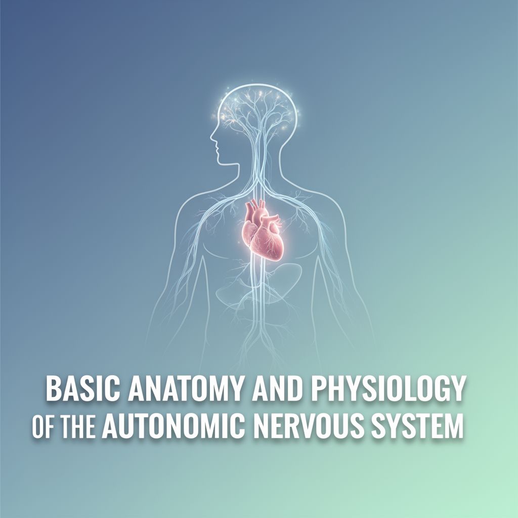
1.2. Autonomic Innervation of the Iris
The Autonomic Nervous System (ANS) controls pupil size:
- PNS Stimulation ✅:
- Neurotransmitter: Acetylcholine (ACh)
- Receptor: Muscarinic (M) receptors on the circular muscle.
- Effect: Pupil constriction (miosis).
- Blocking PNS supply (e.g., with muscarinic antagonists) leads to pupil dilation.
- SNS Stimulation ✅:
- Neurotransmitter: Noradrenaline (NA)
- Receptor: Alpha-1 (α1) adrenoceptors on the radial muscle.
- Effect: Pupil dilation (mydriasis).
- Blocking SNS supply leads to pupil constriction.
1.3. Accommodation of the Lens for Near Vision
The eye's ability to focus on objects at varying distances is called accommodation, primarily for near vision. This involves the ciliary muscle and suspensory ligaments.
- Distant Vision 🔭:
- Ciliary muscle: Relaxed.
- Suspensory ligaments: Taut, stretching the lens thin.
- Lens: Less refractive power needed.
- Near Vision 📖:
- Ciliary muscle: Contracts.
- Suspensory ligaments: Relaxed.
- Lens: Due to its elasticity, it fattens, increasing its refractive power.
- Innervation: This entire process is a PNS action (ACh acting on M receptors).
- Important Note ⚠️: The SNS has no effect on the ciliary muscles or lens accommodation.
1.3.1. Visual Near Point
The visual near point is the closest distance at which an object can be brought into sharp focus.
- Age Effect 📈: The elasticity of the lens decreases with age (due to UV oxidative damage), impairing accommodation. The near point moves further away (e.g., 5-10 cm in a 20-year-old vs. >100 cm in old age).
2. 💊 Drugs and the Eye
2.1. Drug Delivery Challenges
- Blood-Eye Barrier 🛡️: Similar to the blood-brain barrier, it restricts drug access to protect delicate ocular structures.
- Delivery Methods:
- Topical drugs (eye drops) and some oral drugs can reach therapeutic concentrations.
- Injections are sometimes required for effective delivery (e.g., into the vitreous humor).
- Drug Types: Pupil dilators, local anesthetics, pressure regulators, antibiotics, etc.
- Uses: Glaucoma, retinal tears, ocular tumors, cataract procedures, macular degeneration, infections, and more.
2.2. Mechanisms of Drug Action in the Eye
Drugs affecting the eye often target the autonomic nervous system's control over the iris and lens.
- Muscarinic Agonists (e.g., Pilocarpine)
- Action: Mimic PNS effects.
- Effects: Pupil constriction (miosis) and lens accommodation for near vision.
- Muscarinic Antagonists (e.g., Tropicamide, Cyclopentolate)
- Action: Block PNS effects.
- Effects: Pupil dilation (mydriasis) and paralysis of accommodation (cycloplegia), impairing near vision.
- Alpha-1 Adrenergic Agonists (e.g., Phenylephrine)
- Action: Mimic SNS effects on the iris.
- Effects: Pupil dilation (mydriasis).
- Crucially: No effect on lens accommodation, making them useful for ocular examinations where lens focusing is not desired.
3. 🩺 Glaucoma: A Selected Eye Disease
Glaucoma is a serious eye condition characterized by raised intraocular pressure (IOP) that damages the optic nerve, leading to irreversible blindness.
3.1. Overview and Pathophysiology
- Prevalence 📊: Affects ~64 million globally; ~10% of irreversible blindness cases.
- Core Problem: Raised IOP damages the optic nerve.
- Primary Cause: Usually associated with excess aqueous humor.
- Other Factors: Ocular blood flow changes, nerve damage from inflammatory diseases, abnormally low intracranial pressure.
3.2. Types of Glaucoma
- Open-Angle Glaucoma (90% of cases)
- Mechanism: Partially blocked trabecular meshwork (TM), which drains aqueous humor.
- Cause: Accumulation of extracellular matrix (proteins, glycoproteins) forming "plaque material" that impedes outflow.
- Angle-Closure / Narrow-Angle Glaucoma
- Mechanism: Forward bulging of the iris physically blocks the drainage angle between the iris and cornea.
- Risk Factors: Shallow anterior chamber (common in long-sighted individuals), pupil dilation (e.g., in dark rooms or due to drugs).
- Complication: Iris can "stick" to the lens, backing up fluid and pushing the iris further forward, completely blocking drainage.
3.3. Glaucoma Risk Factors
- Age (2x per 10 years after 40y)
- African ethnicity (4x)
- Family history (2-4x)
- Low diastolic perfusion pressure (3x)
- High IOP (e.g., >30mmHg is 40x risk vs. <15mmHg)
- Myopia (1.5-3x)
- Diabetes (1.66x)
3.4. Diagnostic Tests
- Tonometry 💨: Measures IOP (contact or non-contact "puff of air" test).
- Gonioscopy 📐: Measures the angle between the cornea and ciliary body to differentiate open-angle from angle-closure glaucoma.
- Visual Field Test (Perimetry) 🗺️: Checks for missing areas of vision, indicating optic nerve damage.
- Optic Nerve Assessment 📸: Imaging the back of the eye (often requires pupil dilation) to assess the optic disc.
3.5. Glaucoma Treatment Strategies
Treatment aims to reduce IOP by either decreasing aqueous humor production or increasing its outflow.
3.5.1. Decrease Aqueous Production
- α2 Adrenoceptor Agonists (e.g., Brimonidine, Apraclonidine)
- Mechanism: Constrict blood vessels feeding the ciliary body, reducing fluid production. May also increase outflow.
- Side Effects: Red eye, blurred vision.
- β Adrenoceptor Blockers (e.g., Timolol, Levobunolol)
- Mechanism: Antagonize neurotransmission; precise mechanism in glaucoma unclear.
- Side Effects: Blurred vision, irritation, headache.
- Carbonic Anhydrase Inhibitors (e.g., Acetazolamide (oral), Brinzolamide (topical))
- Mechanism: Decrease bicarbonate and sodium movement into aqueous humor, reducing water transport by ~40%.
- Side Effects: Dizziness, dry mouth.
3.5.2. Increase Aqueous Outflow
- Prostaglandin Analogues (e.g., Latanoprost)
- Mechanism: Agonize prostanoid receptors, possibly remodeling the trabecular meshwork and stimulating matrix breakdown.
- Side Effects: Permanent eye color change, longer eyelashes.
- Parasympathomimetics (Muscarinic Agonists) (e.g., Pilocarpine)
- Mechanism: Agonize M3 receptors to contract the ciliary muscle, which opens up the trabecular meshwork, increasing outflow.
- Side Effects: Blurred vision, burning, headache.
- Laser Trabeculoplasty ⚡:
- Procedure: Uses an argon laser to create tiny holes in the trabecular meshwork, allowing greater aqueous outflow.
4. 🧪 Measuring Drug Effects in the Eye (Workshop Examples)
In a practical setting, drug effects can be measured by assessing:
- Pupil diameter (using a pinhole card).
- Light reflex (response to torch light).
- Near-point distance (objective measure of visual acuity).
- Corneal sensation (subjective response to touch with cotton wool).
- Scleral blood vessel constriction (visual assessment of redness).
| Drug | Type | Corneal Sensation | Scleral Vessel Constriction | Pupil Size | Light Reflex | Near Point | | :--------------- | :------------------------------------ | :---------------- | :-------------------------- | :--------- | :----------- | :--------- | | Pilocarpine | Muscarinic Agonist | 0 | 0 | Constrict | Good | Closer | | Tropicamide | Muscarinic Antagonist | 0 | 0 | Dilate | Poor | Further | | Phenylephrine| α1 Adrenoceptor Agonist | 0 | Constrict | Dilate | Good | 0 | | Amethocaine | Local Anesthetic | Less | 0 | 0 | Good | 0 | | Cocaine | Local Anesthetic + NA Reuptake Blocker| Less | Constrict | Dilate | Good | 0 | (Note: '0' indicates no significant change in parameter)
💡 Summary
- PNS Control ✅: Primarily controls both the iris (pupil constriction/miosis) and the lens (accommodation for near vision).
- Iris: Contracts circular muscle.
- Lens: Contracts ciliary body, fattens lens for near vision.
- Aqueous: Opens anterior angle, increases outflow.
- SNS Control ✅: Controls only the iris (pupil dilation/mydriasis), with no direct effect on the lens.
- Iris: Contracts radial muscle.
- Aqueous: May close anterior angle, block outflow.
- Blood Vessels: Constriction.
- Glaucoma ⚠️: A leading cause of irreversible blindness, resulting from raised intraocular pressure (IOP) damaging the optic disc.
- Glaucoma Treatment 💊: Aims to reduce IOP by either decreasing aqueous humor production or increasing its outflow.