This study material has been compiled from a lecture audio transcript and copy-pasted text provided by the user.
📚 The Circulatory and Respiratory Systems: A Comprehensive Study Guide
Introduction
Welcome to this study guide on the human circulatory and respiratory systems! These two vital systems work in close concert to ensure the body's cells receive the oxygen and nutrients they need, while simultaneously removing waste products like carbon dioxide. Understanding their structure and function is fundamental to comprehending human physiology.
🩸 The Circulatory System
The circulatory system is responsible for the movement of materials throughout the body. For complex organisms, where many cells are far from the external environment, this system is indispensable.
1. Components of the Circulatory System
The human circulatory system is a closed system, meaning blood is always contained within vessels. It consists of three primary components:
- Fluid (Blood): The medium in which transported materials are dissolved.
- Network of Tubes (Blood Vessels): The pathways through which the fluid flows.
- Driving Force (Heart): The pump that propels the fluid.
2. The Blood
Blood is a unique connective tissue composed of cells (formed elements) suspended in a liquid matrix called plasma. An average human body contains approximately 5.5 liters of blood.
Functions of Blood ✅
- Transport: Carries respiratory gases (O₂, CO₂), nutrients, cellular wastes, and regulatory substances (enzymes, hormones).
- Regulation: Maintains chemical balance, pH, water content of cells and body fluids, and regulates body temperature.
- Protection:
- Immunity: White blood cells and certain substances protect against disease-causing microorganisms.
- Clotting: The ability to clot prevents excessive fluid loss from wounds, safeguarding the circulatory system.
Composition of Blood 📊
Blood is roughly 55% plasma and 45% formed elements.
a. Plasma
- A clear, straw-colored liquid, primarily 90% water.
- Contains 7% dissolved proteins, along with salts, glucose, amino acids, fatty acids, vitamins, hormones, and cellular wastes.
- Key Plasma Proteins:
- Albumin: Creates an osmotic gradient, regulating plasma diffusion.
- Globulins: Involved in protein transport and body defense against infection (e.g., antibodies).
- Fibrinogen: Essential for blood clotting.
b. Formed Elements
These are the cellular components suspended in plasma.
-
Red Blood Cells (Erythrocytes) 🔴
- Function: Transport oxygen (O₂) from the lungs to tissues and carbon dioxide (CO₂) from tissues back to the lungs.
- Hemoglobin: An iron-containing pigment that gives blood its red color and binds to O₂ and CO₂.
- Characteristics: Lack a nucleus (anucleated) in mature form, allowing more space for hemoglobin. They live for about 120 days.
- Production: Produced in the liver, spleen, and lymph nodes during early embryonic development; after birth, primarily in the bone marrow.
- Anemia: A condition characterized by a deficiency of red blood cells or hemoglobin, leading to reduced oxygen-carrying capacity.
-
White Blood Cells (Leukocytes) ⚪
- Function: Protect the body against infection by bacteria and other microorganisms. They are the body's immune defenders.
- Characteristics: Contain one or more nuclei. Produced by bone marrow and lymphatic tissues.
- Types of White Blood Cells:
- Phagocytes: Engulf pathogens (e.g., neutrophils, macrophages).
- Lymphocytes: Produce antibodies (B cells) and protect against viral infections and destroy cancer cells (T cells).
- Eosinophils: Identify and destroy parasites and assist basophils in allergic responses.
- Basophils: Produce allergic responses (e.g., coughing, sneezing, runny nose).
- Neutrophils: Kill bacteria, fungi, and foreign debris.
- Leukemia: A type of cancer that affects the blood and bone marrow, leading to an overproduction of abnormal white blood cells.
-
Platelets (Thrombocytes) 🩹
- Function: Trigger the blood clotting process.
- Characteristics: Small, round or oval fragments of cells, formed in bone marrow. They consist of cytoplasm surrounded by a cell membrane but lack a nucleus.
3. Blood Vessels
Blood vessels form a vast network of tubes that carry blood throughout the body.
-
Arteries (Distributing Channels) ➡️
- Function: Carry oxygen-rich blood away from the heart to organs and tissues (except pulmonary artery).
- Structure: Possess thick, highly elastic walls composed of connective tissue, muscle tissue, and epithelial tissue. This robust structure allows them to withstand the high pressure of blood pumped directly from the heart.
- Branching: As they penetrate organs and tissues, they progressively branch into smaller vessels called arterioles.
- Valves: Do not contain valves.
- Association: Often accompanied by veins and nerves.
-
Veins (Draining Channels) ⬅️
- Function: Carry oxygen-poor blood back to the heart from body tissues (except pulmonary vein).
- Structure: Have thinner and slightly less elastic walls compared to arteries.
- Branching: The smallest veins are called venules, which collect blood from capillaries.
- Valves: Crucially, veins contain valves that ensure blood flows in one direction only, preventing backflow, especially against gravity.
- Varicose Veins: A condition where veins become enlarged and twisted, often due to faulty valves.
-
Capillaries (Microscopic Vessels) 🕸️
- Function: The primary sites of exchange for oxygen, nutrients, and waste products between blood and tissues.
- Structure: Microscopic vessels that connect arterioles and venules. Their walls consist of a single layer of epithelial cells, making them extremely thin. They are so narrow that red blood cells often pass through in single file. This thinness facilitates rapid diffusion.
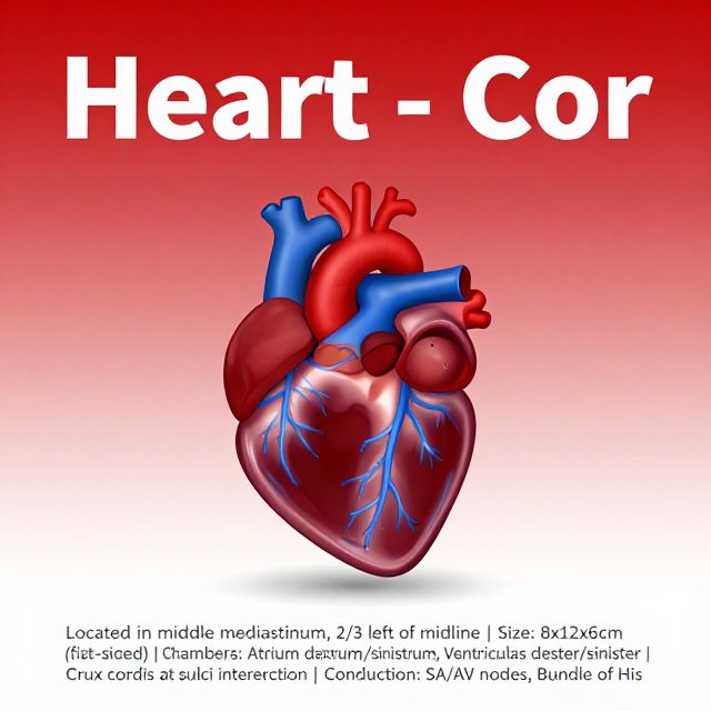
4. The Heart
The heart is a muscular pump, primarily composed of cardiac muscle, which contracts with great force to circulate blood.
Structure of the Heart ❤️
- Cardiac Muscle: Individual cells, each with a nucleus, forming a branching, interlocking network.
- Pericardium: A tough, protective sac of connective tissue filled with fluid, surrounding the outside of the heart.
- Heart Walls (Layers):
- Epicardium: The outermost layer, often containing fat to cushion the heart.
- Myocardium: The thick, muscular middle layer, primarily composed of cardiac muscle. This is the main pumping engine of the heart, with spontaneously contracting fibers that can also conduct electricity.
- Endocardium: The innermost layer, which is thin and smooth, allowing it to stretch as the heart pumps blood.
Internal Chambers and Valves 🚪
The heart is internally divided into four distinct chambers and regulated by four flap-like valves:
- Chambers:
- Atria (Upper Chambers): Two thin-walled chambers (right and left atrium) that receive blood.
- Ventricles (Lower Chambers): Two thick-walled chambers (right and left ventricle) that pump blood out of the heart.
- Septum: A muscular wall that separates the left and right sides of the heart, preventing the mixing of oxygen-rich and oxygen-poor blood.
- Valves: Ensure one-way blood flow.
- Atrioventricular (A-V) Valves: Located between the atria and ventricles.
- Tricuspid Valve: On the right side, between the right atrium and right ventricle.
- Bicuspid (Mitral) Valve: On the left side, between the left atrium and left ventricle.
- Semilunar Valves: Located at the exits of the ventricles into the major arteries.
- Pulmonary Valve: Between the right ventricle and the pulmonary artery.
- Aortic Valve: Between the left ventricle and the aorta.
- Heart Murmurs: Abnormal sounds heard during a heartbeat, often indicating faulty heart valves that allow some blood to flow backward.
- Atrioventricular (A-V) Valves: Located between the atria and ventricles.
Blood Flow through the Heart 🔄
- Right Side: Receives oxygen-poor blood from the body and pumps it to the lungs for oxygenation.
- Left Side: Receives oxygen-rich blood from the lungs and pumps it to the rest of the body.
Functions of the Heart ✅
- Generating Blood Pressure: The force exerted by blood against vessel walls.
- Routing Blood: Separates pulmonary circulation (to lungs) and systemic circulation (to body).
- Ensuring One-Way Blood Flow: Achieved by the precise action of the heart valves.
- Regulating Blood Supply: Adjusts contraction rate and force to match the body's changing metabolic needs.
5. Pathways of Human Circulation
- Pulmonary Circulation: Carries oxygen-poor blood from the heart to the lungs and returns oxygen-rich blood to the heart.
- Systemic Circulation: Carries oxygen-rich blood from the heart to the rest of the body and returns oxygen-poor blood to the heart.
- Coronary Circulation: Specifically supplies blood to the muscle tissue of the heart itself.
- Hepatic-Portal Circulation: Transports blood rich in absorbed nutrients from the digestive tract directly to the liver for processing before it enters general circulation.
- Renal Circulation: Directs blood to and from the kidneys for filtration and waste removal.
6. The Human Lymphatic System
- Function: A complementary system that returns excess fluid and proteins from the intercellular spaces back to the blood. It also plays a crucial role in the body's defense against diseases.
- Lymph Capillaries: One cell layer thick, with flap-like valves, allowing interstitial fluid (lymph) to enter.
- Defense: Lymph nodes filter foreign matter (e.g., cancer cells, bacteria) and produce types of white blood cells (lymphocytes).
🌬️ The Respiratory System
The respiratory system is essential for gas exchange, supporting the metabolic process where glucose and oxygen are converted into carbon dioxide, water, and energy.
1. Requirements for an Efficient Respiratory Surface 💡
For effective gas exchange, a respiratory surface must meet several criteria:
- Thin-walled: To allow rapid diffusion of gases.
- Moist: Oxygen (O₂) and carbon dioxide (CO₂) must be dissolved in solution to diffuse.
- In contact with an environmental source of O₂: To ensure a continuous supply.
- In close contact with the transport system: In multicellular organisms, to efficiently move dissolved materials to and from cells.
2. Components of the Human Respiratory System
The human respiratory system consists of the lungs and a network of air tubes.
-
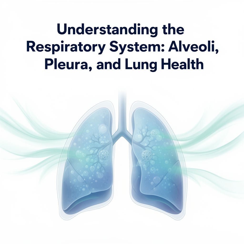
Lungs 🫁
- Location: Occupy a large proportion of the chest (thoracic) cavity.
- Diaphragm: Separated from the abdominal cavity by a muscular sheet called the diaphragm, which aids in breathing.
- Pleura: Each lung is enclosed by a two-layered membrane called the pleura, which reduces friction during breathing.
-
Air Tubes (Respiratory Passageways) 💨
-
Pathway: Air travels through a series of structures:
- Nose: Initial entry point, with long hairs to filter large particles.
- Pharynx (Throat): Common passageway for air and food.
- Trachea (Windpipe): A cartilage-reinforced tube leading to the lungs.
- Bronchi: The trachea branches into two main bronchi, one for each lung.
- Bronchial Tubes: Bronchi further divide into smaller tubes.
- Bronchioles: Even smaller, fine tubes that lack cartilage.
- Alveoli: Tiny air sacs at the end of the bronchioles, which are the primary respiratory surfaces.
-
Protective Features of Air Passageways:
- Ciliated Mucous Membrane: Lines most of the air tubes, secreting mucus to trap particles and moisten the air. Cilia then sweep the mucus and trapped particles upwards, away from the lungs.
- Capillaries: Located beneath the mucous membrane, they warm the inhaled air.
- Adenoids and Tonsils: Lymphoid tissues located in the pharynx, part of the defense system.
- Larynx (Voice Box): Made of cartilage, responsible for voice production.
-
Alveoli: These are the crucial sites for gas exchange. Their walls are only one cell thick, providing an extremely thin barrier for efficient diffusion of O₂ and CO₂ between the air and the blood capillaries surrounding them.
-
3. Phases of Human Respiration reathe
Human respiration involves a sequence of four interconnected phases:
- Breathing (Ventilation): The mechanical process of moving air in and out of the lungs.
- Inhalation: An active process involving muscle contraction (diaphragm and intercostal muscles).
- Exhalation: A passive process, usually occurring as muscles relax.
- External Respiration: The exchange of O₂ and CO₂ between the air in the alveoli of the lungs and the blood in the pulmonary capillaries.
- Circulation: The transport of O₂ from the lungs to the body tissues and CO₂ from the tissues to the lungs via the bloodstream.
- Internal Respiration: The exchange of O₂ and CO₂ between the blood in systemic capillaries and the body cells.
4. Gas Transport in Blood
Gases are transported in the blood in various forms:
-
Oxygen Transport ⬆️
- Primarily transported by hemoglobin (Hb) within red blood cells.
- Hemoglobin binds with oxygen to form oxyhemoglobin (HbO₂).
- Blood Color: Blood low in oxygen (due to deoxyhemoglobin) appears dark red or dull purple. Blood rich in oxygen (due to oxyhemoglobin) appears bright red.
-
Carbon Dioxide Transport ⬇️
- CO₂ is transported in three main ways:
- Bicarbonate Ions (HCO₃⁻): Approximately 70% of CO₂ is transported as bicarbonate ions, formed when CO₂ reacts with water to produce carbonic acid (H₂CO₃), which then dissociates into H⁺ and HCO₃⁻.
- Carbaminohemoglobin (HbCO₂): About 20% of CO₂ binds directly to hemoglobin.
- Dissolved in Plasma: Approximately 10% of CO₂ is transported simply dissolved in the plasma.
- CO₂ is transported in three main ways:
This concludes your study guide on the circulatory and respiratory systems. Remember to review the key terms and processes to solidify your understanding!