This study material has been compiled from the following sources:
- Lecture Audio Transcript: An educational podcast on laboratory diagnosis of gastrointestinal tract infections.
- Personal Notes: Outlines of practical exercises and main topics for the session.
- Copy-pasted Text: Detailed information on gastrointestinal tract infections, pathogens, diagnosis, and therapy.
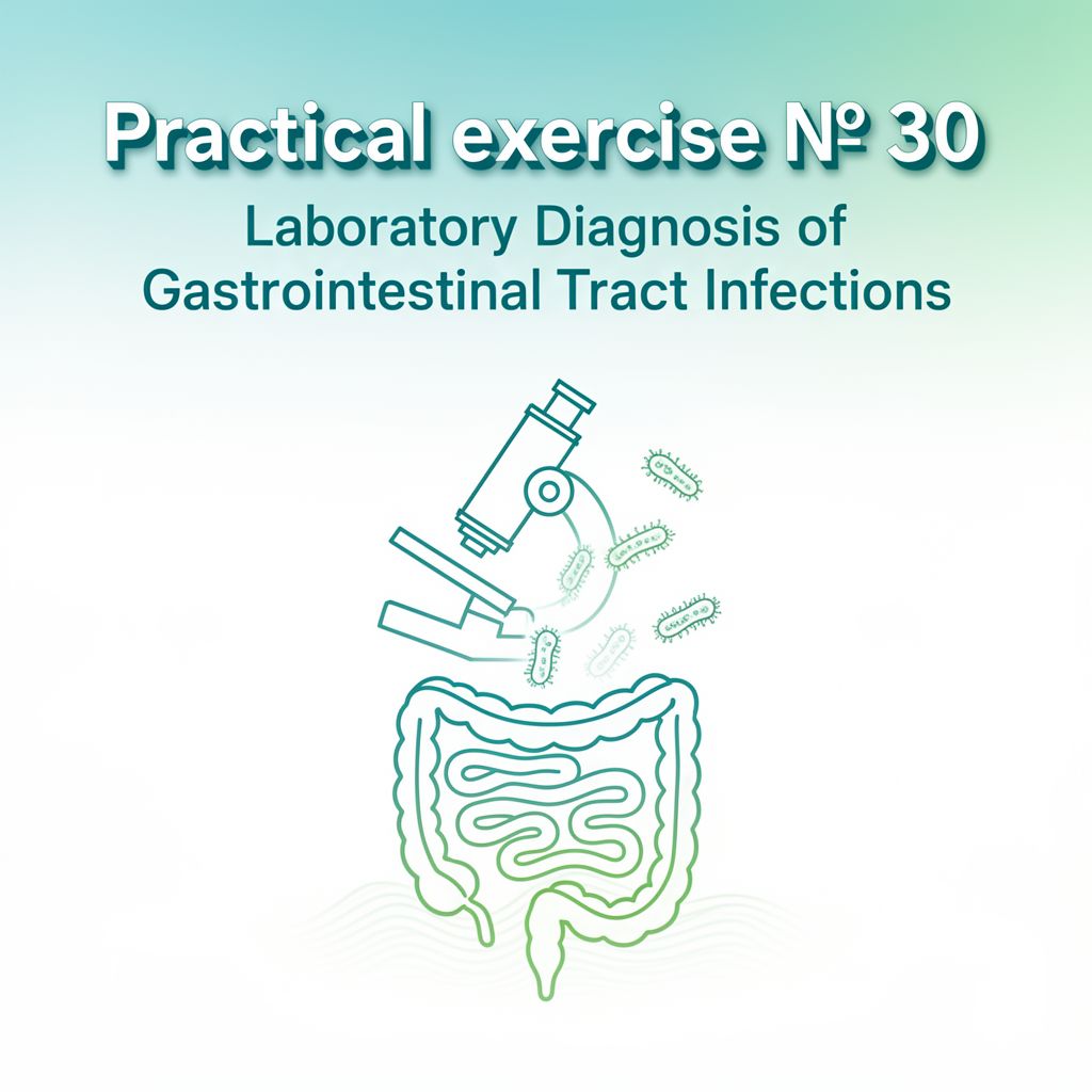
📚 Laboratory Diagnosis of Gastrointestinal Tract Infections
🎯 Introduction to Gastrointestinal Tract Infections (GITIs)
The gastrointestinal tract (GIT) comprises the esophagus, stomach, small intestine (duodenum and ileum), and large intestine (cecum and colon). A wide array of microbial pathogens can infect the GIT, primarily acquired via the fecal-oral route through contaminated food, fluids, or hands.
For an infection to occur, pathogens must be ingested in sufficient numbers or possess attributes to bypass the host defenses of the upper GIT and reach the intestine. Once there, they can cause disease by multiplying and/or producing toxins, or they may invade the intestinal mucosa to reach the lymphatic system or bloodstream.
Symptoms can range from upper GIT issues like nausea, vomiting, cramping, and abdominal pain, to lower GIT involvement with diarrhea, or a combination of both.
📚 Key Definitions
- Gastroenteritis: A syndrome characterized by gastrointestinal symptoms including nausea, vomiting, diarrhea, and abdominal discomfort.
- Diarrhea: Abnormal fecal discharge characterized by frequent and/or fluid stool, usually resulting from small intestine disease and involving increased fluid and electrolyte loss.
- Dysentery: An inflammatory disorder of the GIT often associated with blood and pus in the feces, accompanied by pain, fever, and abdominal cramps, usually resulting from large intestine disease.
- Enterocolitis: Inflammation involving the mucosa of both the small and large intestines.
- Food Poisoning: A type of acute gastroenteritis where the ingestion of a single meal can be identified as the vehicle for infection. Bacteria and bacterial toxins are most commonly implicated.
🦠 Etiological Agents of GITIs
Infectious etiologies include bacterial, viral, and protozoan causes.
1️⃣ Invasive Bacterial Diseases
These pathogens invade the intestinal wall, often leading to inflammatory responses and the presence of fecal leukocytes.
- Salmonella typhi
- Salmonella spp. (non-typhoidal)
- Shigella spp.
- Invasive E. coli (EIEC)
- Yersinia enterocolitica
- Vibrio parahaemolyticus
- Campylobacter spp.
2️⃣ Carrier States
Individuals who harbor pathogens without showing symptoms but can transmit the infection.
- Salmonella typhi
- Non-typhoidal Salmonella
3️⃣ Enterotoxigenic Bacterial Diseases
These pathogens produce toxins that cause symptoms, typically without invading the intestinal wall.
- Enterotoxigenic E. coli (ETEC)
- Verotoxin-producing E. coli (EHEC)
- Vibrio cholerae
- Vibrio parahaemolyticus
- Clostridioides difficile
- Food Poisoning Causes:
- Clostridium perfringens
- Clostridium botulinum
- Staphylococcus aureus
- Bacillus cereus
4️⃣ Viral Agents
- Rotavirus (common cause of infantile diarrhea)
- Norwalk virus
5️⃣ Parasitic Agents
- Entamoeba histolytica
- Giardia lamblia
- Cryptosporidium
🧪 Laboratory Diagnosis of Infectious Gastroenteritis
1️⃣ Collection and Transport of Specimens
Laboratory confirmation typically involves recovering pathogenic microorganisms from various specimens.
- Stool (Feces):
- Collected in a clean, waxed cardboard or plastic container.
- Minimum volume: 1 teaspoon (approx. 5 mL) for most procedures.
- Preferably collect parts containing abnormal material (pus, mucus).
- If delay > 2 hours is anticipated, place in transport medium (e.g., Cary-Blair medium for optimal viability).
- Rectal Swabs:
- May be necessary for Shigella spp. recovery if stool is unavailable.
- ⚠️ Note: Not suitable for detecting parasites, toxins, or viral antigens.
- Other Specimens:
- Blood Cultures: Essential for febrile gastroenteritis patients, especially in typhoid fever where other sources might be negative.
- Bone Marrow Cultures, Urine, CSF, Material from Rose Spots: Can be cultured in cases of typhoid fever.
- Duodenal Aspirates: Useful for diagnosing duodenal parasites (G. lamblia) and isolating S. typhi from carriers or acute typhoid fever patients.
- Gastric Biopsy Material: For detecting H. pylori in peptic ulcer disease.
- Material from Mucosal Lesions: Obtained during proctoscopy or sigmoidoscopy for C. difficile detection.
- Serum Samples: For Widal test in typhoid fever and septicemic Salmonella infections.
2️⃣ Direct Detection of Pathogens in Stool
a) Microscopic Examinations
- Gram Stain: Generally not useful for stool, as most pathogens cannot be differentiated from the normal Gram-negative bowel flora.
- Methylene Blue Staining Wet Mount for Fecal Leukocytes (WBCs):
- ✅ Fecal leukocytes indicate inflammatory disease and provide clues to etiology.
- ✅ Present in bacterial infections that invade the intestinal wall (e.g., Shigella, invasive E. coli).
- ❌ Not seen in viral gastroenteritis, parasitic diarrhea, enterotoxigenic bacterial diarrhea, or Salmonella carrier state.
- Wet Mount:
- 💡 Fastest method for detecting intestinal parasites.
- Used with darkfield microscopy for observing darting motility and curved forms of Campylobacter.
- Hanging Drop Mount: Used in endemic areas to recognize the comma-shaped appearance and motility of Vibrio cholerae.
b) ELISA Procedures and Latex Agglutination Tests
- Available for direct detection of rotavirus.
c) Nucleic Acid Probe Technologies
- Commercially available for detection of Campylobacter spp. and rotavirus.
3️⃣ Cultures
Stools for routine cultures are examined for "enteric pathogens" like Salmonella and Shigella.
a) Routine Cultures
Typically involve a sequence of media:
- Enrichment Broth: (e.g., GN or Selenite broth) to promote growth of pathogens while suppressing normal flora.
- Differential Medium: (e.g., MacConkey's, EMB-Levine's agar) to differentiate non-lactose-fermenting (often pathogens) from lactose-fermenting enteric bacteria.
- Selective Medium: (e.g., SS agar) to inhibit the growth of most enterobacteria, allowing Salmonella and Shigella to be detected.
b) Special Enrichment Techniques and/or Media
Required for specific fastidious or slow-growing pathogens:
- Yersinia enterocolitica: "Cold enrichment" of fecal cultures; SS agar or special medium (CIN agar).
- Vibrio cholerae: Alkaline peptone water, alkaline agar, TCBS agar.
- Campylobacter jejuni: Campy-blood agar or Skirrow's selective medium, incubated at 40-42°C in microaerophilic conditions for 48-72 hours.
- Helicobacter pylori: Campy-blood agar or Skirrow's selective medium, incubated at 35°C microaerophilically in high humidity for up to 7 days.
4️⃣ Specific Tests for Identification
Once microorganisms are isolated, further tests are needed for definitive identification.
a) Biochemical Tests
- Rapid and conventional biochemical tests are used to identify isolated microorganisms based on their metabolic characteristics.
b) Rapid Slide Agglutination Tests
- Definitive identification of isolated pathogens (e.g., EPEC, Shigella spp., Salmonella spp., Vibrio cholerae) is accomplished using rapid slide agglutination tests with specific antisera.
5️⃣ Serologic Tests for Enteric Pathogens
- Serum samples are used for tests like the Widal test in typhoid fever and septicemic Salmonella infections to detect antibodies against specific pathogens.
6️⃣ Antimicrobial Susceptibility Tests
- Antimicrobial susceptibility testing on isolated pathogens should be done routinely to guide appropriate treatment.
💊 Therapy for Gastrointestinal Tract Infections
-
Supportive Therapy:
- The cornerstone of treatment for most infectious diarrheas.
- Fluid Replacement: Maintaining adequate hydration is the most important aspect, especially in infants and children.
- Antispasmodics and antiemetics may also be used.
-
Antimicrobial Therapy:
- Necessary: In enteric fevers and septicemic Salmonella infections.
- Shortens Symptom Duration: In Shigella, Campylobacter, Y. enterocolitica, and V. cholerae infections.
- ⚠️ Caution: Can prolong the carrier state of uncomplicated Salmonella gastroenteritis.
- Antimicrobial susceptibility testing is crucial to ensure effective treatment.
🧪 Practical Tasks for Laboratory Diagnosis
- Examination of wet mount preparations from a stool.
- Observation of growth characteristics of enteric pathogens.
- Reading biochemical tests for identification.
- Performing slide agglutination tests for detection of EPEC, Shigella spp., Salmonella spp., and Vibrio cholerae.
- Interpretation of results from disk-diffusion susceptibility tests.