Muscles of the Lower Limb: Leg Compartments - kapak
Bilim#anatomy#lower limb#leg muscles#fibularis

Muscles of the Lower Limb: Leg Compartments

Explore the anatomy of the leg muscles, categorized by their compartments: lateral, anterior, and posterior. Learn about the origin, insertion, innervation, and actions of key muscles.

rana_lJanuary 21, 2026 ~15 dk toplam
01

Flash Kartlar

25 kart

Karta tıklayarak çevir. ← → ile gez, ⎵ ile çevir.

1 / 25
Tüm kartları metin olarak gör
  1. 1. What are the main functions of the leg muscles?

    The muscles of the leg are crucial for controlling foot movements like flexion, extension, inversion, and eversion. These actions provide stability to the lower limb during movements at the knee and hip joints, enabling various forms of locomotion and balance. Understanding these muscles involves categorizing them into distinct compartments.

  2. 2. Name the three primary compartments of the leg discussed.

    The three primary compartments of the leg are the lateral, anterior, and posterior compartments. Each compartment contains specific muscles with distinct functions, origins, insertions, and innervations, allowing for organized and efficient movement of the foot and toes. These divisions help in understanding their coordinated actions.

  3. 3. Which muscles are found in the lateral compartment of the leg?

    The lateral compartment of the leg primarily houses two muscles: the Fibularis longus and the Fibularis brevis. These muscles are mainly responsible for eversion of the foot and assist in plantar flexion, playing a key role in foot stability and movement. They are crucial for maintaining balance on uneven surfaces.

  4. 4. What is the primary action of the muscles in the lateral compartment?

    The primary action of the muscles in the lateral compartment (Fibularis longus and Fibularis brevis) is eversion of the foot at the subtalar joint. They also assist in plantar flexion at the talocrural joint, contributing to the overall stability and movement of the ankle and foot. This combined action helps in adapting the foot to various terrains.

  5. 5. What is the innervation for both Fibularis longus and Fibularis brevis?

    Both the Fibularis longus and Fibularis brevis muscles are innervated by the superficial fibular nerve. This nerve typically originates from spinal segments L5 and S1, ensuring coordinated control over the eversion and plantar flexion actions of these lateral compartment muscles. Damage to this nerve can impair foot eversion.

  6. 6. Describe the origin and insertion of the Fibularis longus.

    The Fibularis longus originates from the head and proximal two-thirds of the lateral surface of the fibula, partially arising from the intermuscular septa. It inserts onto the plantar side of the medial cuneiform and the base of the first metatarsal. This long path allows it to support the transverse arch of the foot.

  7. 7. What is a unique function of the Fibularis longus compared to Fibularis brevis?

    While both perform plantar flexion and eversion, the Fibularis longus uniquely supports the transverse arch of the foot. Its long tendon crosses the sole of the foot to insert on the medial cuneiform and first metatarsal, providing crucial stability to the arch. This distinguishes its role in foot mechanics.

  8. 8. What is the main function of the anterior compartment muscles of the leg?

    The muscles of the anterior compartment are primarily responsible for dorsiflexion of the foot at the talocrural joint. They also contribute to the extension of the toes, playing a vital role in lifting the foot during the swing phase of gait and preventing toe drag. This action is essential for normal walking.

  9. 9. Name the four significant muscles found in the anterior compartment.

    The anterior compartment includes four significant muscles: the Tibialis anterior, Extensor hallucis longus, Extensor digitorum longus, and Fibularis tertius. These muscles work synergistically to achieve dorsiflexion and toe extension, allowing for precise control of foot and toe movements. They are crucial for activities like walking and running.

  10. 10. Which nerve innervates all muscles of the anterior compartment?

    All muscles of the anterior compartment (Tibialis anterior, Extensor hallucis longus, Extensor digitorum longus, and Fibularis tertius) are innervated by the deep fibular nerve. This nerve typically originates from spinal segments L4 and L5, ensuring coordinated control over dorsiflexion and toe extension. Injury to this nerve can result in 'foot drop'.

  11. 11. What are the primary actions of the Tibialis anterior?

    The Tibialis anterior primarily performs dorsiflexion at the talocrural joint and inversion (supination) at the subtalar joint. It is a strong muscle crucial for lifting the foot and maintaining the medial longitudinal arch, especially during walking and running. Its dual action makes it vital for foot stability and movement.

  12. 12. Where does the Extensor hallucis longus insert?

    The Extensor hallucis longus inserts at the dorsal aponeurosis at the base of the distal phalanx of the first toe. This specific insertion allows it to extend the big toe, as well as contribute to dorsiflexion of the foot and active eversion/inversion depending on foot position. It plays a key role in fine control of the great toe.

  13. 13. What is the main action of the Extensor digitorum longus?

    The Extensor digitorum longus primarily extends the metatarsophalangeal and interphalangeal joints of the second to fifth toes. It also contributes to dorsiflexion at the talocrural joint and eversion at the subtalar joint, working to lift and splay the lesser toes. This muscle is important for clearing the toes during gait.

  14. 14. Which anterior compartment muscle originates from the distal fibula and inserts on the fifth metatarsal?

    The Fibularis tertius originates from the anterior border of the distal fibula and inserts onto the base of the fifth metatarsal. It is considered a part of the Extensor digitorum longus and contributes to dorsiflexion at the talocrural joint and eversion at the subtalar joint. Its presence can be variable among individuals.

  15. 15. How does the Extensor hallucis longus contribute to foot movement beyond toe extension?

    Beyond extending the big toe, the Extensor hallucis longus also performs dorsiflexion at the talocrural joint. It can actively contribute to both eversion and inversion at the subtalar joint, depending on the initial position of the foot, making it versatile in foot positioning. This adaptability is important for balance and gait.

  16. 16. Compare the insertion points of Tibialis anterior and Fibularis tertius.

    The Tibialis anterior inserts onto the medial and plantar surface of the medial cuneiform and the medial base of the first metatarsal. In contrast, the Fibularis tertius inserts onto the base of the fifth metatarsal. This difference reflects their roles in inversion (Tibialis anterior) versus eversion (Fibularis tertius) of the foot, contributing to different aspects of foot stability.

  17. 17. How is the posterior compartment of the leg divided?

    The posterior compartment of the leg is divided into superficial and deep flexors. These two groups are separated by the transverse intermuscular septum, allowing for distinct functional roles in plantar flexion, inversion, and toe flexion. This anatomical separation helps organize the complex actions of the posterior leg muscles.

  18. 18. Name the three muscles that form the "triceps surae."

    The "triceps surae" refers to the superficial flexors of the posterior compartment, which include the Gastrocnemius, Soleus, and Plantaris muscles. These three muscles converge to form the calcaneal (Achilles) tendon, a powerful plantar flexor unit. They are essential for propulsion during walking and running.

  19. 19. What is the primary innervation for the superficial flexors of the posterior compartment?

    The primary innervation for the superficial flexors (Gastrocnemius, Soleus, and Plantaris) of the posterior compartment is the tibial nerve. This nerve typically originates from spinal segments S1 and S2, coordinating their powerful plantar flexion actions. This innervation ensures efficient movement and strength in the ankle.

  20. 20. Describe the origin and primary action of the Gastrocnemius.

    The Gastrocnemius has two heads: the medial head originates from the superior posterior part of the medial femoral condyle, and the lateral head originates from the lateral surface of the lateral femoral condyle. It is a powerful plantar flexor at the talocrural joint when the knee is extended, and it also contributes to knee flexion. This makes it a biarticular muscle.

  21. 21. How does the Soleus muscle differ from the Gastrocnemius in terms of origin and action?

    The Soleus originates from the head and neck of the fibula and the soleal line of the tibia, unlike the Gastrocnemius which originates from the femur. While both are powerful plantar flexors, the Soleus is a pure ankle plantar flexor, active regardless of knee position, whereas Gastrocnemius's plantar flexion is strongest with an extended knee. This difference is important for sustained plantar flexion.

  22. 22. What is the functional significance of the Plantaris muscle?

    The Plantaris muscle originates from the lateral epicondyle of the femur and inserts into the calcaneal tuberosity. While also innervated by the tibial nerve, its action is considered negligible. It may assist the Gastrocnemius in plantar flexion, but its primary role is often considered proprioceptive or as a vestigial muscle, sometimes mistaken for a nerve.

  23. 23. What are the general functions of the deep flexors in the posterior compartment?

    The deep flexors of the posterior compartment play crucial roles in foot inversion, plantar flexion, and flexion of the toes. They are essential for fine motor control of the foot and toes, as well as supporting the arches of the foot. These muscles contribute significantly to balance and intricate foot movements.

  24. 24. Name the four muscles found in the deep posterior compartment.

    The four muscles found in the deep posterior compartment are the Tibialis posterior, Flexor digitorum longus, Flexor hallucis longus, and Popliteus. These muscles are vital for various foot and toe movements, as well as knee unlocking. They are arranged deep to the superficial flexors.

  25. 25. Which deep posterior compartment muscle has a broad insertion and supports foot arches?

    The Tibialis posterior has a broad insertion, attaching to the navicular tuberosity, the medial, intermediate, and lateral cuneiforms, and the bases of the second to fourth metatarsals. Its actions include plantar flexion, inversion, and crucially, supporting both the longitudinal and transverse arches of the foot. This makes it a key stabilizer of the foot.

02

Bilgini Test Et

15 soru

Çoktan seçmeli sorularla öğrendiklerini ölç. Cevap + açıklama.

Soru 1 / 15Skor: 0

Which of the following muscles is primarily responsible for supporting the transverse arch of the foot?

03

Detaylı Özet

5 dk okuma

Tüm konuyu derinlemesine, başlık başlık.

📚 Lower Limb Muscles: Leg Compartments Study Guide

This study material is compiled from a lecture audio transcript and copy-pasted text, providing a comprehensive overview of the muscles in the leg compartments.


Introduction to Leg Muscle Compartments

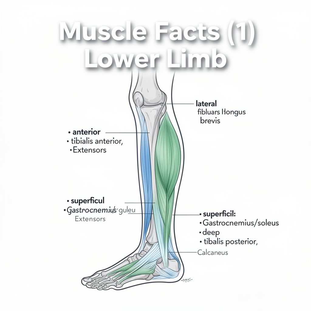
The muscles of the leg are fundamental for controlling foot movements, including flexion, extension, inversion, and eversion. These actions are crucial for maintaining stability in the lower limb during movements at both the knee and hip joints. Understanding these muscles involves categorizing them into distinct compartments, each with specific functions, origins, insertions, and innervations. This guide will detail the lateral, anterior, and posterior compartments of the leg.


1. Lateral Compartment Muscles

The lateral compartment of the leg primarily contains muscles responsible for eversion of the foot and assisting in plantar flexion. These muscles are key for dynamic stability during walking and running.

1.1. Fibularis Longus (Peroneus Longus)

  • 📚 Origin: Head and proximal two-thirds of the lateral surface of the fibula, partially from intermuscular septa.
  • 📚 Insertion: Plantar side of the medial cuneiform and base of the first metatarsal.
  • 📚 Innervation: Superficial fibular nerve (L5, S1).
  • ✅ Actions:
    • Talocrural joint: Plantar flexion.
    • Subtalar joint: Eversion (pronation).
    • 💡 Supports the transverse arch of the foot.

1.2. Fibularis Brevis (Peroneus Brevis)

  • 📚 Origin: Distal half of the lateral surface of the fibula, intermuscular septa.
  • 📚 Insertion: Tuberosity at the base of the fifth metatarsal (with an occasional division to the dorsal aponeurosis of the fifth toe).
  • 📚 Innervation: Superficial fibular nerve (L5, S1).
  • ✅ Actions:
    • Talocrural joint: Plantar flexion.
    • Subtalar joint: Eversion (pronation).

2. Anterior Compartment Muscles

The anterior compartment muscles are primarily responsible for dorsiflexion of the foot and extension of the toes. They are essential for lifting the foot during the swing phase of gait.

2.1. Tibialis Anterior

  • 📚 Origin: Upper two-thirds of the lateral surface of the tibia, interosseous membrane, and superficial crural fascia.
  • 📚 Insertion: Medial and plantar surface of the medial cuneiform, and medial base of the first metatarsal.
  • 📚 Innervation: Deep fibular nerve (L4, L5).
  • ✅ Actions:
    • Talocrural joint: Dorsiflexion.
    • Subtalar joint: Inversion (supination).

2.2. Extensor Hallucis Longus

  • 📚 Origin: Middle third of the medial surface of the fibula and interosseous membrane.
  • 📚 Insertion: Dorsal aponeurosis at the base of the distal phalanx of the first toe.
  • 📚 Innervation: Deep fibular nerve (L4, L5).
  • ✅ Actions:
    • Talocrural joint: Dorsiflexion.
    • Subtalar joint: Active in both eversion and inversion (pronation/supination), depending on the initial position of the foot.
    • Extends the metatarsophalangeal (MTP) and interphalangeal (IP) joints of the big toe.

2.3. Extensor Digitorum Longus

  • 📚 Origin: Head and medial surface of the fibula, lateral condyle of the tibia, and interosseous membrane.
  • 📚 Insertion: Dorsal aponeuroses at the bases of the distal phalanges of the second to fifth toes.
  • 📚 Innervation: Deep fibular nerve (L4, L5).
  • ✅ Actions:
    • Talocrural joint: Dorsiflexion.
    • Subtalar joint: Eversion (pronation).
    • Extends the MTP and IP joints of the second to fifth toes.

2.4. Fibularis Tertius (Peroneus Tertius)

  • 📚 Origin: Distal fibula (anterior border).
  • 📚 Insertion: Base of the fifth metatarsal.
  • 📚 Innervation: Deep fibular nerve (L4, L5).
  • ✅ Actions:
    • Talocrural joint: Dorsiflexion.
    • Subtalar joint: Eversion (pronation).

3. Posterior Compartment Muscles

The posterior compartment of the leg is divided into two groups: superficial and deep flexors. These groups are separated by the transverse intermuscular septum.

3.1. Superficial Flexors (Triceps Surae)

These muscles form the bulk of the calf and are powerful plantar flexors of the foot.

3.1.1. Gastrocnemius

  • 📚 Origin:
    • Medial head: Superior posterior part of the medial femoral condyle.
    • Lateral head: Lateral surface of the lateral femoral condyle.
  • 📚 Insertion: Calcaneal tuberosity via the calcaneal (Achilles') tendon.
  • 📚 Innervation: Tibial nerve (S1, S2).
  • ✅ Actions:
    • Talocrural joint: Powerful plantar flexion (especially when the knee is extended).
    • Knee joint: Flexion.

3.1.2. Soleus

  • 📚 Origin: Head and neck of the fibula, and the soleal line of the tibia via a tendinous arch.
  • 📚 Insertion: Calcaneal tuberosity via the calcaneal (Achilles') tendon.
  • 📚 Innervation: Tibial nerve (S1, S2).
  • ✅ Actions:
    • Talocrural joint: Plantar flexion.

3.1.3. Plantaris

  • 📚 Origin: Lateral epicondyle of the femur, proximal to the lateral head of the Gastrocnemius.
  • 📚 Insertion: Calcaneal tuberosity.
  • 📚 Innervation: Tibial nerve (S1, S2).
  • ✅ Actions:
    • Negligible; may assist Gastrocnemius in plantar flexion.

3.2. Deep Flexors

These muscles are crucial for foot inversion, plantar flexion, and toe flexion, and play a significant role in supporting the arches of the foot.

3.2.1. Tibialis Posterior

  • 📚 Origin: Interosseous membrane, adjacent borders of the tibia and fibula.
  • 📚 Insertion: Navicular tuberosity, medial, intermediate, and lateral cuneiforms, and bases of the second to fourth metatarsals.
  • 📚 Innervation: Tibial nerve (L4, L5).
  • ✅ Actions:
    • Talocrural joint: Plantar flexion.
    • Subtalar joint: Inversion (supination).
    • 💡 Supports the longitudinal and transverse arches of the foot.

3.2.2. Flexor Digitorum Longus

  • 📚 Origin: Middle third of the posterior surface of the tibia.
  • 📚 Insertion: Bases of the second to fifth distal phalanges.
  • 📚 Innervation: Tibial nerve (L5-S2).
  • ✅ Actions:
    • Talocrural joint: Plantar flexion.
    • Subtalar joint: Inversion (supination).
    • MTP and IP joints of the second to fifth toes: Plantar flexion.

3.2.3. Flexor Hallucis Longus

  • 📚 Origin: Distal two-thirds of the posterior surface of the fibula, adjacent interosseous membrane.
  • 📚 Insertion: Base of the first distal phalanx.
  • 📚 Innervation: Tibial nerve (L4-S1).
  • ✅ Actions:
    • Talocrural joint: Plantar flexion.
    • Subtalar joint: Inversion (supination).
    • MTP and IP joints of the first toe: Plantar flexion.
    • 💡 Supports the medial longitudinal arch.

3.2.4. Popliteus

  • 📚 Origin: Lateral femoral condyle, posterior horn of the lateral meniscus.
  • 📚 Insertion: Posterior tibial surface (above the origin of the Soleus).
  • 📚 Innervation: Tibial nerve (L4, L5).
  • ✅ Actions:
    • Knee joint: Flexes and "unlocks" the knee by externally rotating the femur on the fixed tibia.

Kendi çalışma materyalini oluştur

PDF, YouTube videosu veya herhangi bir konuyu dakikalar içinde podcast, özet, flash kart ve quiz'e dönüştür. 1.000.000+ kullanıcı tercih ediyor.

Sıradaki Konular

Tümünü keşfet
Forearm Muscles: Anatomy and Clinical Relevance

Forearm Muscles: Anatomy and Clinical Relevance

An in-depth look at the anterior and posterior compartments of the forearm, detailing muscle origins, insertions, innervations, actions, and clinical conditions like lateral epicondylitis.

10 dk Özet 25 15
The Musculoskeletal System: Structure, Function, and Locomotion

The Musculoskeletal System: Structure, Function, and Locomotion

Explore the intricate musculoskeletal system, its components, functions, and the mechanisms of locomotion in various organisms, with a detailed focus on the human body.

Özet 25 15 Görsel
Fundamental Biological Principles and Nutrition

Fundamental Biological Principles and Nutrition

An academic summary covering biological organization from cells to organ systems, key cellular processes, major organ system functions, and essential nutritional components with their identification tests.

5 dk Özet 25 15
Skeletal Muscle Structure and Neuromuscular Junction

Skeletal Muscle Structure and Neuromuscular Junction

This audio summary provides an academic overview of skeletal muscle terminology, morphology, filament and sarcomere structure, innervation, and the intricate process of excitation-contraction coupling.

7 dk Özet 18 12
Neuron Types and Nervous System Structures

Neuron Types and Nervous System Structures

This summary explores the diverse morphology and classification of neurons, their fundamental components, synaptic connections, and the crucial role of glial cells and peripheral nervous system structures.

6 dk Özet 25 15
The Visual Pathway, Reflexes, and Eye Movements

The Visual Pathway, Reflexes, and Eye Movements

An academic overview of the visual pathway from retina to cortex, including visual field processing, magnocellular and parvocellular streams, light and accommodation reflexes, and conjugate eye movements.

8 dk Özet 25 15
The Anatomy and Physiology of the Human Heart

The Anatomy and Physiology of the Human Heart

Explore the intricate structure and function of the human heart, covering its location, chambers, valves, and the electrical conduction system that drives its rhythmic beat.

Özet 25 15
The Vestibular System and Auditory Pathway

The Vestibular System and Auditory Pathway

An academic overview of the vestibular system's role in balance and spatial orientation, and the auditory pathway's function in sound perception, detailing their anatomical structures and neural processing.

8 dk Özet 25 15