📚 Study Material: Forearm Muscles of the Upper Limb
Source Information: This study material has been compiled from a lecture audio transcript and accompanying anatomical figures and tables (Fig. 26.12, Table 26.2, Fig. 26.14, Clinical box 26.4, Table 26.3, Fig. 26.16, Table 26.4).
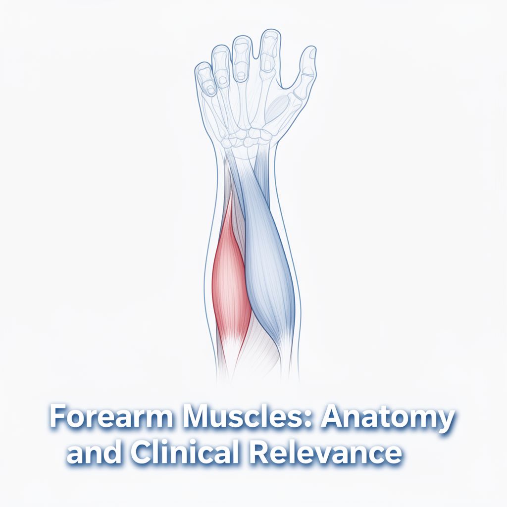
🧠 Introduction to Forearm Muscles
This guide provides a detailed overview of the complex muscular structure of the forearm, focusing on both the anterior and posterior compartments. We will explore the origin, insertion, innervation, and actions of each muscle, along with a common clinical condition known as Lateral Epicondylitis (Tennis Elbow). Understanding these muscles is crucial for comprehending human anatomy and muscle function.
1️⃣ Anterior Compartment of the Forearm
The anterior compartment muscles are primarily responsible for flexion and pronation movements of the forearm and wrist. They are organized into three layers: superficial, intermediate, and deep.
A. Superficial Muscles
These muscles generally originate from the medial epicondyle of the humerus.
- 1. Pronator Teres
- 📚 Origin: Humeral head (medial epicondyle of humerus), Ulnar head (coronoid process of ulna)
- 📚 Insertion: Lateral radius (distal to supinator insertion)
- 📚 Innervation: Median nerve (C6, C7)
- 📚 Action: Elbow: weak flexion; Forearm: pronation
- 2. Flexor Carpi Radialis
- 📚 Origin: Medial epicondyle of humerus
- 📚 Insertion: Base of 2nd metacarpal (sometimes 3rd metacarpal)
- 📚 Innervation: Median nerve (C7, C8)
- 📚 Action: Wrist: flexion and abduction (radial deviation) of hand; Elbow: weak flexion
- 3. Palmaris Longus
- 📚 Origin: Medial epicondyle of humerus
- 📚 Insertion: Palmar aponeurosis
- 📚 Innervation: Median nerve (C7, C8)
- 📚 Action: Wrist: flexion; tightens palmar aponeurosis; Elbow: weak flexion
- 4. Flexor Carpi Ulnaris
- 📚 Origin: Humeral head (medial epicondyle), Ulnar head (olecranon)
- 📚 Insertion: Pisiform, hook of hamate, base of 5th metacarpal
- 📚 Innervation: Ulnar nerve (C7-T1)
- 📚 Action: Wrist: flexion and adduction (ulnar deviation) of hand; Elbow: weak flexion
B. Intermediate Muscles
- 1. Flexor Digitorum Superficialis
- 📚 Origin: Humeral-ulnar head (medial epicondyle of humerus and coronoid process of ulna), Radial head (upper half of anterior border of radius)
- 📚 Insertion: Sides of middle phalanges of 2nd to 5th digits
- 📚 Innervation: Median nerve (C8, T1)
- 📚 Action: Wrist, MCP, and PIP joints of 2nd to 5th digits: flexion; Elbow: weak flexion
C. Deep Muscles
- 1. Flexor Digitorum Profundus
- 📚 Origin: Ulna (proximal two-thirds of flexor surface) and interosseous membrane
- 📚 Insertion: Distal phalanges of 2nd to 5th digits (palmar surface)
- 📚 Innervation: Median nerve (C8, T1, radial half of fingers 2 and 3); Ulnar nerve (C8, T1, ulnar half of fingers 4 and 5)
- 📚 Action: Wrist, MCP, PIP, and DIP joints of 2nd to 5th digits: flexion
- 2. Flexor Pollicis Longus
- 📚 Origin: Radius (mid-anterior surface) and adjacent interosseous membrane
- 📚 Insertion: Distal phalanx of thumb (palmar surface)
- 📚 Innervation: Median nerve (C8, T1)
- 📚 Action: Carpometacarpal joint of thumb: flexion; MCP and IP joints of thumb: flexion; Wrist: flexion and abduction (radial deviation) of hand
- 3. Pronator Quadratus
- 📚 Origin: Distal quarter of ulna (anterior surface)
- 📚 Insertion: Distal quarter of radius (anterior surface)
- 📚 Innervation: Median nerve (C8, T1)
- 📚 Action: Hand: pronation; Distal radioulnar joint: stabilization
2️⃣ Posterior Compartment of the Forearm
The posterior compartment muscles are primarily responsible for extension and supination movements of the forearm and wrist. They are organized into radialis muscles, superficial, and deep layers.
A. Radialis Muscles
These muscles are located on the radial side of the forearm.
- 1. Brachioradialis
- 📚 Origin: Distal humerus (lateral surface), lateral intermuscular septum
- 📚 Insertion: Styloid process of the radius
- 📚 Innervation: Radial nerve (C5, C6)
- 📚 Action: Elbow: flexion; Forearm: semipronation
- 2. Extensor Carpi Radialis Longus
- 📚 Origin: Lateral supracondylar ridge of distal humerus, lateral intermuscular septum
- 📚 Insertion: Base of 2nd metacarpal
- 📚 Innervation: Radial nerve (C6, C7)
- 📚 Action: Elbow: weak flexion; Wrist: extension and abduction
- 3. Extensor Carpi Radialis Brevis
- 📚 Origin: Lateral epicondyle of humerus
- 📚 Insertion: Base of 3rd metacarpal
- 📚 Innervation: Radial nerve (C7, C8)
- 📚 Action: Wrist: extension and abduction
B. Superficial Muscles
These muscles generally originate from the lateral epicondyle of the humerus.
- 1. Extensor Digitorum
- 📚 Origin: Common head (lateral epicondyle of humerus)
- 📚 Insertion: Dorsal digital expansion of 2nd to 5th digits
- 📚 Innervation: Radial nerve (C7, C8)
- 📚 Action: Wrist: extension; MCP, PIP, and DIP joints of 2nd to 5th digits: extension/abduction of fingers
- 2. Extensor Digiti Minimi
- 📚 Origin: Common head (lateral epicondyle of humerus)
- 📚 Insertion: Dorsal digital expansion of 5th digit
- 📚 Innervation: Radial nerve (C7, C8)
- 📚 Action: Wrist: extension, ulnar abduction of hand; MCP, PIP, and DIP joints of 5th digit: extension and abduction of 5th digit
- 3. Extensor Carpi Ulnaris
- 📚 Origin: Common head (lateral epicondyle of humerus), Ulnar head (dorsal surface)
- 📚 Insertion: Base of 5th metacarpal
- 📚 Innervation: Radial nerve (C7, C8)
- 📚 Action: Wrist: extension, adduction (ulnar deviation) of hand
C. Deep Muscles
- 1. Supinator
- 📚 Origin: Olecranon, lateral epicondyle of humerus, radial collateral ligament, annular ligament of radius
- 📚 Insertion: Radius (between radial tuberosity and insertion of pronator teres)
- 📚 Innervation: Radial nerve (C6, C7)
- 📚 Action: Radioulnar joints: supination
- 2. Abductor Pollicis Longus
- 📚 Origin: Radius and ulna (dorsal surfaces, interosseous membrane)
- 📚 Insertion: Base of 1st metacarpal
- 📚 Innervation: Radial nerve (C7, C8)
- 📚 Action: Radiocarpal joint: abduction of the hand; Carpometacarpal joint of thumb: abduction
- 3. Extensor Pollicis Brevis
- 📚 Origin: Radius (posterior surface) and interosseous membrane
- 📚 Insertion: Base of proximal phalanx of thumb
- 📚 Innervation: Radial nerve (C7, C8)
- 📚 Action: Radiocarpal joint: abduction (radial deviation) of hand; Carpometacarpal and MCP joints of thumb: extension
- 4. Extensor Pollicis Longus
- 📚 Origin: Ulna (posterior surface) and interosseous membrane
- 📚 Insertion: Base of distal phalanx of thumb
- 📚 Innervation: Radial nerve (C7, C8)
- 📚 Action: Wrist: extension and abduction (radial deviation) of hand; Carpometacarpal joint of thumb: adduction; MCP and IP joints of thumb: extension
- 5. Extensor Indicis
- 📚 Origin: Ulna (posterior surface) and interosseous membrane
- 📚 Insertion: Posterior digital expansion of 2nd digit
- 📚 Innervation: Radial nerve (C7, C8)
- 📚 Action: Wrist: extension; MCP, PIP, and DIP joints of 2nd digit: extension
🏥 Clinical Relevance: Lateral Epicondylitis (Tennis Elbow)
⚠️ Lateral epicondylitis, commonly known as tennis elbow, is a condition affecting the extensor muscles and tendons of the forearm that attach to the lateral epicondyle of the humerus.
- Affected Tendon: The tendon most frequently involved is that of the Extensor Carpi Radialis Brevis. This muscle helps stabilize the wrist when the elbow is extended.
- Mechanism:
- Overuse leads to weakening of the tendon.
- Microscopic tears form where the tendon attaches to the lateral epicondyle.
- This results in inflammation and pain.
- 💡 Evidence suggests inflammation can extend along the tendon to the periosteum of the lateral epicondyle.
- Affected Population:
- While named "tennis elbow," athletes are actually a minority of those affected.
- The condition is often referred to as "lateral elbow syndrome."
- Individuals whose activities require repetitive and vigorous use of forearm muscles are particularly prone. This includes:
- Painters
- Plumbers
- Carpenters
- Auto workers
- Cooks
- Butchers
- Signs and Symptoms:
- Pain with wrist extension against resistance.
- Point tenderness or burning sensation on the lateral epicondyle.
- Weak grip strength.
- Symptoms are intensified with forearm activity.
📊 Key Abbreviations
- DIP: Distal Interphalangeal joint
- IP: Interphalangeal joint
- MCP: Metacarpophalangeal joint
- PIP: Proximal Interphalangeal joint