This study material has been compiled from various sources, including copy-pasted text and a lecture audio transcript, to provide a comprehensive overview of Riboflavin (Vitamin B2) and Niacin (Vitamin B3).
Essential B Vitamins: Riboflavin (B2) & Niacin (B3)
Introduction
This guide explores two crucial B vitamins: Riboflavin (Vitamin B2) and Niacin (Vitamin B3). We will cover their digestion, absorption, transport, storage, vital functions, metabolic pathways, and the clinical implications of their deficiency and toxicity. Understanding these vitamins is fundamental to comprehending cellular energy metabolism and overall health.
1. Riboflavin (Vitamin B2) 💡
Riboflavin, also known as Vitamin B2, is an essential water-soluble vitamin that plays a critical role in numerous metabolic processes.
1.1. Digestion, Absorption, Transport, and Storage
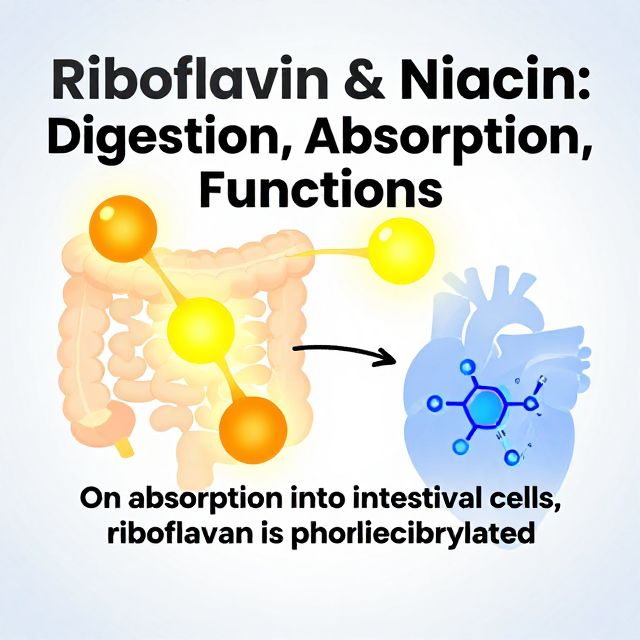
- Absorption ✅
- Upon absorption into intestinal cells, riboflavin is phosphorylated to form Flavin Mononucleotide (FMN).
- This reaction is catalyzed by the enzyme flavokinase and requires ATP.
- Transport 📊
- In the blood, flavins are found as riboflavin (approximately 50%), FMN (10%), and FAD (40%).
- These forms are transported in plasma by various proteins, including albumin, fibrinogen, and globulins (primarily immunoglobulins).
- Free riboflavin is the form that traverses most cell membranes, primarily through a carrier-mediated process involving a riboflavin-binding protein.
- Storage 📚
- Riboflavin is stored in small quantities across various tissues.
- Highest concentrations are found in the liver, kidneys, and heart.
- The body typically stores enough riboflavin to meet needs for about 2 to 6 weeks.
- Within cells, riboflavin is converted into its coenzyme forms:
- FMN (Flavin Mononucleotide): Major intracellular form (60–95%).
- FAD (Flavin Adenine Dinucleotide): (5–20%).
- This conversion is facilitated by flavokinase and FAD synthetase, enzymes widely distributed in tissues like the liver, spleen, small intestine, kidneys, and heart.
1.2. Functions and Mechanisms of Action
Riboflavin's primary function is as a precursor to the coenzymes FMN and FAD, which are integral to many oxidative enzyme systems, known as flavoproteins.
- Coenzyme Roles ✅
- FMN and FAD function as coenzymes for a wide variety of oxidative enzyme systems, remaining bound to enzymes during oxidation-reduction reactions.
- Flavins act as oxidizing agents due to their ability to accept a pair of hydrogen atoms.
- Electron Transport Chain: Flavoproteins play a crucial role in the electron transport chain, facilitating energy production.
- Vitamin B6 Metabolism: FMN is essential for pyridoxine phosphate oxidase, which converts pyridoxamine phosphate (PMP) and pyridoxine phosphate (PNP) to pyridoxal phosphate (PLP), the active form of Vitamin B6.
- Oxidative Decarboxylation: FAD serves as an intermediate electron carrier in the oxidative decarboxylation of pyruvate and α-ketoglutarate, leading to NADH production.
- Succinate Dehydrogenase: This FAD flavoprotein removes electrons from succinic acid to form fumarate, producing FADH2, which then passes electrons into the electron transport chain via coenzyme Q.
- Fatty Acid Beta-Oxidation: Acyl-CoA dehydrogenases, crucial for fatty acid breakdown, require FAD.
- Folate Synthesis: FADH2 is required for the synthesis of the active form of folate, 5-methyl THF.
- Niacin Synthesis: FAD is needed for kynureninase monooxygenase, an enzyme involved in a step of niacin synthesis from tryptophan.
- Oxidases: FAD acts as a coenzyme for oxidases like xanthine oxidase and aldehyde oxidase, transferring electrons directly to oxygen to form hydrogen peroxide.
- Glutathione Reductase: This FAD-dependent enzyme is vital for reducing oxidized glutathione (GSSG) to its reduced form (GSH), protecting cells from oxidative stress.
1.3. Metabolism and Excretion
- Riboflavin and its metabolites are primarily excreted in the urine.
- Free riboflavin (not protein-bound) is filtered by the glomerulus and excreted.
- Most riboflavin (60–70%) is excreted intact in the urine.
- Characteristic Urine Color: Oral ingestion of riboflavin, especially in supplement quantities (e.g., 1.7 mg), can cause urine to deepen from light yellow to a brighter, orangish-yellow due to its fluorescent properties. This effect is often noticeable within hours.
1.4. Toxicity ⚠️
- Toxicity associated with large oral doses of riboflavin has not been reported.
- Consequently, no Tolerable Upper Intake Level (UL) for riboflavin has been established.
1.5. Deficiency: Ariboflavinosis
- An acute deficiency, known as ariboflavinosis, rarely occurs in isolation and is often accompanied by other nutrient deficits.
- Symptoms 📝:
- Cheilosis: Painful lesions or fissures on the outside of the lips.
- Angular Stomatitis: Lesions at the corners of the mouth.
- Glossitis: Inflammation of the tongue.
1.6. Requirements
- Recommended Dietary Allowances (RDAs) for adults:
- Men: 1.3 mg/day
- Women: 1.1 mg/day
1.7. Assessment of Nutrient Status 📊
- The most sensitive method involves measuring the activity of the FAD-dependent enzyme erythrocyte glutathione reductase.
- In riboflavin deficiency, the activity of this enzyme is limited.
- Activity Coefficients (AC) are determined with and without added FAD:
- AC < 1.2: Acceptable riboflavin status.
- AC of 1.2 to 1.4: Low riboflavin status.
- AC > 1.4: Suggests riboflavin deficiency.
- Other indicators:
- Cellular riboflavin concentrations < 10 μg/dL.
- Urinary riboflavin excretion < 19 μg/g creatinine (without recent intake) or < 40 μg per day.
2. Niacin (Vitamin B3) 💡
Niacin, or Vitamin B3, is a crucial B vitamin known for its role in energy metabolism and its historical link to the deficiency disease pellagra.
2.1. Discovery and Forms
- Discovery: Niacin was discovered through its deficiency disorder, pellagra, in humans, and a similar condition called "black tongue" in dogs. It was once called the "anti–black tongue factor."
- Pellagra was prevalent in the southeastern United States in the early 1900s due to corn-based diets, which contain niacin in an unavailable form.
- Elvehjem isolated the vitamin in 1937, demonstrating its ability to cure pellagra and black tongue.
- Forms 📚:
- Niacin is a generic term for nicotinic acid and nicotinamide (also called niacinamide), both providing vitamin activity.
- A third form, nicotinamide riboside, is less commonly used clinically.
- Structurally: Nicotinic acid is pyridine 3-carboxylic acid, while nicotinamide is nicotinic acid amide.
- Essential Chemical Features:
- Pyridine nucleus substituted with a β-carboxylic acid or corresponding amine.
- Pyridine nitrogen must undergo reversible oxidation/reduction.
- Pyridine carbons adjacent to the nuclear nitrogen atom must be open.
2.2. Dietary Sources and Biosynthesis
- Dietary Sources ✅:
- Best sources: Most fish and meats.
- Other sources: Enriched cereals and bread products, whole grains, fortified cereals, seeds, legumes.
- Lesser amounts: Green vegetables, milk.
- Coffee and tea also contain niacin; trigonelline in coffee is converted to niacin by heat.
- Forms in Food: In animal foods, niacin is mainly nicotinamide and its nucleotides (NAD, NADP). In plant foods, it's mainly nicotinic acid. Niacin in food is stable to cooking and storage.
- Biosynthesis from Tryptophan 🧬:
- Niacin can be synthesized in the liver and other tissues from the amino acid tryptophan.
- Approximately 1 mg of niacin is produced from 60 mg of dietary tryptophan.
- This pathway requires other nutrients: riboflavin (FAD), vitamin B6 (PLP), and iron. Deficiencies in these can impair niacin synthesis.
2.3. Digestion, Absorption, Transport, and Storage
- Digestion 1️⃣:
- Digestion of NAD and NADP is required for niacin absorption.
- A pyrophosphatase hydrolyzes phosphate from NADP.
- NAD is hydrolyzed by glycohydrolase, releasing free nicotinamide.
- Absorption 2️⃣:
- Nicotinamide and nicotinic acid can be absorbed in the stomach, but are more readily absorbed in the small intestine.
- Primarily by sodium-dependent, carrier-mediated (facilitated) diffusion.
- At high pharmacological doses (3–4 g), niacin is absorbed almost completely by passive diffusion.
- Transport 3️⃣:
- In plasma, niacin is found primarily as nicotinamide, but also as nicotinic acid (up to one-third bound to plasma proteins).
- Nicotinamide and nicotinic acid move across cell membranes by simple diffusion.
- However, nicotinic acid transport into kidney tubules and red blood cells requires a carrier, and uptake into the brain is energy-dependent.
- Conversion & Storage 📚:
- Nicotinamide is the primary precursor for NAD (Nicotinamide Adenine Dinucleotide) synthesis in all tissues.
- Nicotinic acid can also synthesize NAD, mainly in the liver.
- NAD is phosphorylated by NAD kinase (using ATP) to generate NADP (Nicotinamide Adenine Dinucleotide Phosphate).
- These reactions are reversible.
- The vitamin is trapped within cells as NAD or NADP.
- Intracellular NAD concentrations typically predominate over NADP.
- In the liver, excess niacin and tryptophan are converted to NAD, stored in small amounts not bound to enzymes.
- NAD is primarily found in its oxidized form (NAD+), while NADP is mainly in its reduced form (NADPH).
2.4. Functions and Mechanisms of Action
Niacin's coenzymes, NAD and NADP, are vital for approximately 200 enzymes, primarily dehydrogenases, acting as hydrogen donors or electron acceptors.
- Coenzyme Roles ✅:
- NADH (from NAD): Major role is to transfer electrons from metabolic intermediates through the electron transport chain, producing ATP.
- Oxidative Reactions: Glycolysis, oxidative decarboxylation of pyruvate to acetyl-CoA, oxidation of acetyl-CoA in the TCA cycle, β-oxidation of fatty acids, oxidation of ethanol.
- Also required by aldehyde dehydrogenase for catabolism of vitamin B6 (pyridoxal to pyridoxic acid).
- NADPH (from NADP): Acts as a reducing agent in many biosynthetic pathways.
- Reductive Biosynthesis: Fatty acid synthesis, cholesterol and steroid hormone synthesis, proline synthesis, deoxyribonucleotide (DNA precursors) synthesis.
- Antioxidant Systems: Glutathione, vitamin C, and thioredoxin regeneration.
- Folate Coenzyme Synthesis.
- NADPH is produced in the pentose phosphate pathway and the mitochondrial membrane malate-aspartate shuttle.
- NADH (from NAD): Major role is to transfer electrons from metabolic intermediates through the electron transport chain, producing ATP.
- Nonredox Roles 🧬:
- NAD acts as a donor of adenosine diphosphate ribose (ADP-ribose) for post-translational modification of proteins and formation of cyclic ADP-ribose.
- These ADP-ribosylation reactions control cellular processes such as:
- DNA repair, replication, and transcription.
- G-protein activity.
- Chromatin structure.
- Intracellular calcium signaling.
2.5. Metabolism and Excretion
- NAD and NADP undergo degradation in cells by glycohydrolase to form ADP-ribose and nicotinamide.
- The released nicotinamide is then methylated and oxidized in the liver into various products, which are excreted in the urine.
- Primary Metabolites 📊:
- N'-methyl nicotinamide (NMN): ~20–30% of niacin metabolites.
- N'-methyl 2-pyridone 5-carboxamide (2-pyridone): ~40–60%.
- Small amounts of N'-methyl 4-pyridone carboxamide (4-pyridone).
- Nicotinic acid is mainly metabolized to N'-methyl-nicotinic acid.
- Little free nicotinic acid or nicotinamide is excreted, as both are actively reabsorbed from the glomerular filtrate.
2.6. Toxicity ⚠️
- Due to the vasodilatory effects (e.g., "niacin flush") associated with supplemental niacin, a Tolerable Upper Intake Level (UL) for adults has been set.
- UL: 35 mg/day for niacin (both nicotinic acid and nicotinamide) from supplements and fortified foods.
2.7. Deficiency: Pellagra
- A deficiency of niacin results in pellagra (from Italian, "pelle agra" meaning "rough skin").
- Pellagra is classically characterized by the "Four Ds" 📝:
- Dermatitis: Skin lesions, often symmetrical and appearing in sun-exposed areas.
- Dementia: Neurological symptoms including confusion, memory loss, and depression.
- Diarrhea: Gastrointestinal disturbances.
- Death: If untreated.
- Gastrointestinal Manifestations: Glossitis, cheilosis, angular stomatitis, nausea, vomiting, and diarrhea.
- Treatment: Typically requires about 500 mg of nicotinamide daily for several weeks.
2.8. Requirements
- Niacin recommendations account for niacin derived from tryptophan, using Niacin Equivalents (NE).
- 1 mg of niacin is estimated to be produced from 60 mg of dietary tryptophan.
- Recommended Dietary Allowances (RDAs) for adults:
- Men: 16 mg NE/day
- Women: 14 mg NE/day
- Estimated Average Requirements (EARs): Men 12 mg, Women 11 mg. (RDAs are typically 30% higher than EARs).
2.9. Assessment of Nutrient Status 📊
- Niacin status is primarily assessed by measuring the urinary excretion of its metabolites.
- Urinary N'-methyl nicotinamide:
- < 0.8 mg/day or < 0.5 mg/g creatinine: Suggestive of poor (deficient) niacin status.
- 0.5 to 1.59 mg/g creatinine: Suggests marginal niacin status.
-
1.6 mg/g creatinine: Reflects adequate status.