Skeletal Muscle Physiology: Structure, Function, and Regulation - kapak
Bilim#skeletal muscle#muscle physiology#sarcomere#actin

Skeletal Muscle Physiology: Structure, Function, and Regulation

This summary explores the molecular structure of skeletal muscle, neuromuscular transmission, excitation-contraction coupling, ATP-dependent contraction, and mechanisms of force regulation and fiber type classification.

aleyblgnMarch 29, 2026 ~23 dk toplam
01

Sesli Özet

6 dakika

Konuyu otobüste, koşarken, yolda dinleyerek öğren.

Sesli Özet

Skeletal Muscle Physiology: Structure, Function, and Regulation

0:006:04
02

Flash Kartlar

25 kart

Karta tıklayarak çevir. ← → ile gez, ⎵ ile çevir.

1 / 25
Tüm kartları metin olarak gör
  1. 1. What is the smallest contractile unit of skeletal muscle?

    The smallest contractile unit of skeletal muscle is the muscle fiber. It is a multinucleated, elongated cell whose organized architecture allows for significant mechanical force generation. Each muscle fiber contains myofibrils, which are composed of repeating sarcomeres.

  2. 2. Describe the basic structure of a sarcomere.

    A sarcomere is the basic contractile unit of a myofibril, consisting of interdigitating thin and thick myofilaments. Six thin filaments, primarily actin, surround each thick filament, primarily myosin, in a hexagonal arrangement. This precise organization is crucial for muscle contraction.

  3. 3. What are the primary proteins that make up thin filaments in skeletal muscle?

    Thin filaments are primarily composed of actin, specifically F-actin, which is a double-stranded alpha-helical polymer of actin molecules. Associated with F-actin are the regulatory proteins tropomyosin and troponin, which play key roles in controlling muscle contraction by regulating the availability of myosin-binding sites on actin.

  4. 4. What are the primary proteins that make up thick filaments in skeletal muscle?

    Thick filaments are bipolar assemblies of multiple myosin II molecules. Each myosin II molecule consists of two intertwined heavy chains and two pairs of light chains. The myosin heads, extending from the thick filament, interact with actin during muscle contraction, forming cross-bridges.

  5. 5. Define a neuromuscular junction.

    A neuromuscular junction is a specialized chemical synapse formed between a motor nerve terminal and a skeletal muscle fiber. This specific region on the muscle fiber is often referred to as the end plate, where neural signals are transmitted to initiate muscle contraction through the release of neurotransmitters.

  6. 6. What is a motor unit?

    A motor unit comprises all the muscle fibers that are innervated by a single motor neuron. When a motor neuron fires an action potential, all the muscle fibers within its motor unit contract simultaneously. This coordinated contraction allows for graded control of muscle force.

  7. 7. Which neurotransmitter is released at the neuromuscular junction, and what type of receptor does it activate?

    Acetylcholine is the neurotransmitter released at the neuromuscular junction. It activates nicotinic acetylcholine receptors located on the muscle fiber's end plate. This activation leads to the production of an excitatory end-plate current, which initiates muscle depolarization and subsequent contraction.

  8. 8. Explain the underlying cause of Myasthenia Gravis.

    Myasthenia Gravis is an autoimmune disorder where the body produces antibodies against its own nicotinic acetylcholine receptors at the neuromuscular junction. This leads to a reduction in the number of functional receptors on the muscle fiber's surface. Consequently, the transmission of nerve impulses to muscle fibers is impaired.

  9. 9. How does Myasthenia Gravis affect muscle function?

    Due to the reduced number of acetylcholine receptors, the end-plate potential generated at the neuromuscular junction is diminished. This makes it harder for the muscle fiber to reach its threshold for depolarization and generate an action potential. The result is muscle weakness and fatigability, especially with repetitive use.

  10. 10. How are acetylcholinesterase inhibitors used to treat Myasthenia Gravis?

    Acetylcholinesterase inhibitors, such as neostigmine, are used to treat Myasthenia Gravis by prolonging the action of acetylcholine in the synaptic cleft. By inhibiting the enzyme that breaks down acetylcholine, these drugs allow the neurotransmitter to remain active longer. This partially compensates for the loss of receptors, improving muscle strength.

  11. 11. What is the role of T-tubules in skeletal muscle contraction?

    Transverse tubules, or T-tubules, are invaginations of the sarcolemma that penetrate deep into the muscle fiber. They play a crucial role in propagating action potentials from the sarcolemma into the interior of the muscle cell. This ensures rapid and synchronous activation of all myofibrils, leading to a coordinated contraction.

  12. 12. What is the ultimate signal for muscle contraction, and where is it released from?

    The ultimate signal for muscle contraction is a rise in intracellular calcium concentration ([Ca2+]i). This calcium is released from the sarcoplasmic reticulum, a specialized endoplasmic reticulum found in muscle cells. This release occurs in response to an action potential propagating along the T-tubules.

  13. 13. Define excitation-contraction (EC) coupling.

    Excitation-contraction (EC) coupling is the physiological process that links the electrical excitation of the muscle fiber (action potential) to the mechanical event of muscle contraction. Specifically, it describes the sequence of events from T-tubule depolarization to the increase in intracellular calcium concentration that ultimately initiates cross-bridge cycling.

  14. 14. What are L-type calcium channels (DHP receptors) and what is their function in EC coupling?

    L-type calcium channels, also known as dihydropyridine (DHP) receptors, are voltage-gated channels located in the T-tubule membrane. In skeletal muscle, they primarily act as voltage sensors. Upon T-tubule depolarization, they undergo conformational changes that directly trigger calcium release from the sarcoplasmic reticulum.

  15. 15. How do DHP receptors interact with ryanodine receptors to release calcium?

    Upon T-tubule depolarization, the conformational change in the DHP receptors (L-type calcium channels) directly induces a change in the calcium release channel, known as the ryanodine receptor, located in the sarcoplasmic reticulum membrane. This mechanical coupling facilitates the rapid release of calcium into the cytoplasm, initiating contraction.

  16. 16. How does increased intracellular calcium trigger muscle contraction?

    Increased intracellular calcium ([Ca2+]i) triggers muscle contraction by removing the inhibition of cross-bridge cycling. Calcium binds to regulatory proteins, specifically troponin C, which then causes a conformational change in tropomyosin. This shift exposes the myosin-binding sites on actin filaments, allowing myosin heads to attach and initiate contraction.

  17. 17. Explain the role of troponin C in skeletal muscle contraction.

    Troponin C is a component of the troponin complex that binds calcium. In skeletal muscle, it has two pairs of Ca2+-binding sites: two high-affinity sites that are always occupied, and two low-affinity sites that bind and release Ca2+ as intracellular calcium concentration fluctuates. This binding regulates the interaction between actin and myosin by controlling tropomyosin's position.

  18. 18. What is the primary energy source for the cross-bridge cycle?

    The cross-bridge cycle, which drives muscle contraction, converts the energy from ATP hydrolysis into mechanical energy. ATP binds to the myosin head, allowing it to detach from actin, and its hydrolysis provides the energy for the myosin head to re-cock and bind to a new site on actin. This continuous cycle requires a constant supply of ATP.

  19. 19. How do muscles regenerate ATP during exercise?

    During exercise, muscles regenerate ATP using various sources. Initially, stored ATP and creatine phosphate are used for immediate energy. For sustained activity, muscle glycogen and blood glucose are utilized through glycolysis and oxidative phosphorylation. At rest, aerobic respiration of fatty acids is the primary method for ATP regeneration.

  20. 20. What are the two main uses of ATP in muscle contraction and relaxation?

    ATP powers two critical processes in muscle function: first, it provides the energy for the cross-bridge movement during contraction, allowing myosin heads to detach from actin and re-cock. Second, ATP is required for the active re-uptake of calcium into the sarcoplasmic reticulum by SERCA pumps, which is essential for muscle relaxation.

  21. 21. How is muscle contraction terminated?

    Muscle contraction is terminated by the active re-uptake of calcium into the sarcoplasmic reticulum. This process removes calcium from the cytoplasm, causing it to dissociate from troponin C. As a result, tropomyosin re-covers the myosin-binding sites on actin, preventing further cross-bridge formation and leading to muscle relaxation.

  22. 22. What is the role of calsequestrin in the sarcoplasmic reticulum?

    Calsequestrin is a calcium-binding protein located within the sarcoplasmic reticulum lumen, particularly beneath the triad junction. Its role is to buffer the high intracellular calcium concentration during re-uptake. By binding calcium, it enhances the sarcoplasmic reticulum's calcium storage capacity, facilitating efficient calcium removal from the cytoplasm and promoting relaxation.

  23. 23. How does a single muscle fiber increase its force of contraction over time?

    In a single muscle fiber, the force of contraction increases by summing multiple twitches over time. If successive action potentials arrive before the muscle has completely relaxed, the subsequent contractions add to the previous ones. This phenomenon, known as temporal summation, can lead to a greater overall force, or even tetanus if stimulation is rapid enough.

  24. 24. How does a whole muscle increase its force of contraction?

    In a whole muscle, force increases by summing the contractions of multiple muscle fibers. The central nervous system regulates muscle force by recruiting more motor units into the contracting pool, a process called multiple-fiber summation or spatial summation. This mechanism allows for a graded and stronger muscle contraction, adapting to the required force.

  25. 25. How does the central nervous system ensure smooth, non-tetanic contraction for fine motor control?

    The central nervous system achieves smooth, non-tetanic contraction for fine motor control by activating individual motor units asynchronously. This means that different motor units are stimulated at different times, preventing all fibers from reaching tetanus simultaneously. This asynchronous activation allows for a more continuous and controlled force output, essential for precise movements.

03

Bilgini Test Et

15 soru

Çoktan seçmeli sorularla öğrendiklerini ölç. Cevap + açıklama.

Soru 1 / 15Skor: 0

What is the smallest contractile unit of skeletal muscle?

04

Detaylı Özet

7 dk okuma

Tüm konuyu derinlemesine, başlık başlık.

📚 Skeletal Muscle Physiology: A Comprehensive Study Guide

Source Information: This study material has been compiled and organized from various sources, including copy-pasted text and a lecture audio transcript on Skeletal Muscle Physiology.


Introduction to Skeletal Muscle Physiology

Skeletal muscle is essential for movement, posture, and heat generation. Its function relies on a highly organized molecular structure, precise neural control, and efficient energy utilization. This guide will explore the molecular architecture of skeletal muscle, the process of neuromuscular transmission, excitation-contraction coupling, the energetics of muscle contraction, and the mechanisms regulating muscle force, including different muscle fiber types.


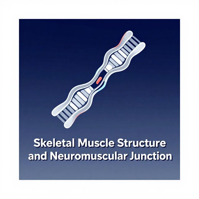
I. Molecular Structure of Skeletal Muscle

Skeletal muscle exhibits a hierarchical organization, allowing it to generate significant mechanical force.

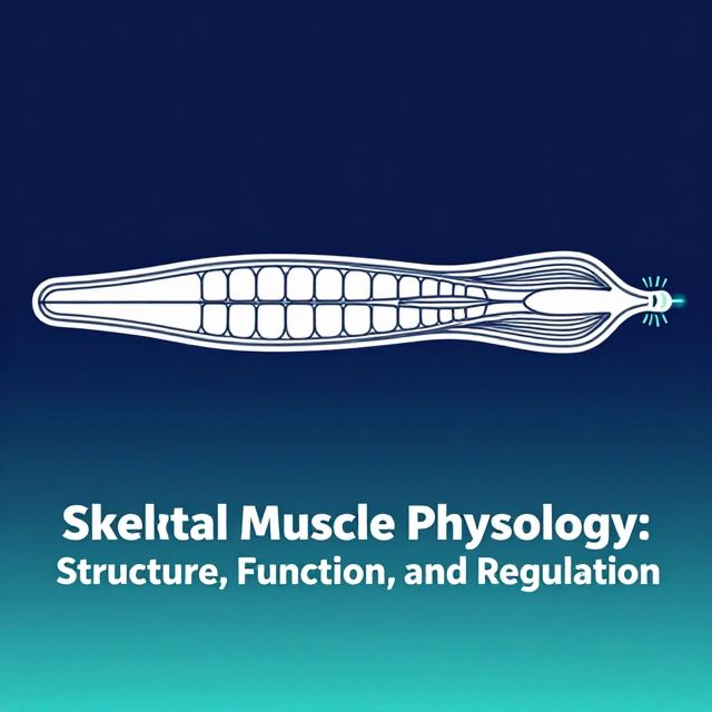
A. Muscle Fiber and Myofibrils

  • Muscle Fiber (Myofiber): The smallest contractile unit of skeletal muscle. It is a multinucleated, elongated cell. ✅
  • Myofibril: Each muscle fiber contains numerous myofibrils, which are essentially end-to-end chains of repeating contractile units called sarcomeres.

B. Myofilaments: Thin and Thick

Within each sarcomere, smaller interdigitating filaments, known as myofilaments, are responsible for contraction.

1. Thin Filaments (Actin)

  • Composition: The backbone of the thin filament is a double-stranded α-helical polymer of actin molecules, known as filamentous actin (F-actin). Each helical turn consists of 13 individual actin monomers. 📚
  • Associated Proteins: F-actin is associated with two crucial regulatory proteins:
    • Tropomyosin: A long, fibrous protein that, in a relaxed state, covers the myosin-binding sites on actin.
    • Troponin: A complex of three proteins (Troponin I, T, and C) that regulates the position of tropomyosin. Troponin C is the calcium-binding component.

2. Thick Filaments (Myosin)

  • Composition: Thick filaments are bipolar assemblies of multiple myosin II molecules. Each myosin II molecule is composed of:
    • Two intertwined heavy chains.
    • Two alkali (or essential) light chains.
    • Two regulatory light chains.
  • Arrangement: Six thin filaments (actin) surround each thick filament (myosin) in a tightly packed hexagonal array, allowing for optimal overlap during contraction.

II. Neuromuscular Junction and Transmission

The precise control of skeletal muscle contraction begins with neural signaling at specialized synapses.

A. Motor Unit

  • Definition: A motor unit comprises a single motor neuron and all the muscle fibers it innervates. ✅
  • Function: When a motor neuron fires an action potential, all muscle fibers within its motor unit contract simultaneously.

B. Acetylcholine and Receptors

  • Neuromuscular Junction (NMJ): This is the chemical synapse between peripheral motor nerve terminals and skeletal muscle fibers, also known as the end plate. 💡
  • Neurotransmitter: Acetylcholine (ACh) is released from the motor neuron terminal.
  • Receptors: ACh activates nicotinic acetylcholine receptors (nAChRs) on the muscle fiber membrane, producing an excitatory end-plate current (EPC) and subsequently an end-plate potential (EPP).

C. Clinical Correlation: Myasthenia Gravis

  • Pathology: Myasthenia Gravis is an autoimmune disorder caused by the presence of antibodies that attack and reduce the number of ACh receptors on the muscle end plate. ⚠️
  • Symptoms: Characterized by skeletal muscle weakness and fatigability.
  • Mechanism: The reduced number of ACh receptors leads to a diminished EPP, making it harder to depolarize the muscle membrane to threshold and generate action potentials.
  • Treatment: Acetylcholinesterase (AChE) inhibitors (e.g., neostigmine) prevent the degradation of ACh, prolonging its action at the NMJ and partially compensating for the receptor loss.

III. Excitation-Contraction (EC) Coupling

EC coupling is the process by which an electrical signal (excitation) is converted into a mechanical response (contraction).

A. Role of Action Potentials and T-Tubules

  • Signal Propagation: Action potentials propagate from the sarcolemma (muscle cell membrane) into the interior of the muscle fiber along the transverse tubule (T-tubule) network. ✅
  • Ultimate Signal: The ultimate intracellular signal that triggers and sustains muscle contraction is a rise in intracellular calcium concentration ([Ca2+]i).

B. Calcium Release from Sarcoplasmic Reticulum

  • Storage: Ca2+ is stored in the sarcoplasmic reticulum (SR), an intracellular Ca2+ storage reservoir.
  • Mechanism:
    1. T-tubule Depolarization: The action potential propagating into the T-tubules depolarizes the triad region (where T-tubules meet SR).
    2. DHP Receptor Activation: This depolarization activates L-type Ca2+ channels, also known as dihydropyridine (DHP) receptors, located in the T-tubule membrane. These act as voltage sensors.
    3. Ryanodine Receptor Activation: Conformational changes in the DHP receptors induce conformational changes in the Ca2+ release channel (ryanodine receptor, RyR) located in the SR membrane.
    4. Ca2+ Release: This opens the RyR, leading to a rapid release of Ca2+ from the SR into the cytoplasm.

C. Mechanism of Contraction Initiation (Calcium's Role)

  • Inhibition Removal: The increase in [Ca2+]i triggers contraction by removing the inhibition of cross-bridge cycling.
  • Regulatory Proteins: Ca2+ exerts its effect by binding to regulatory proteins, not directly to contractile proteins.
    • In the absence of Ca2+, tropomyosin covers the myosin-binding sites on actin, preventing actin-myosin interaction.
    • In skeletal muscle, troponin C has two pairs of Ca2+-binding sites:
      • High-affinity sites: Always occupied by Ca2+ or Mg2+ under physiological conditions.
      • Low-affinity sites: These bind and release Ca2+ as [Ca2+]i rises and falls in the sarcoplasm. When Ca2+ binds to these sites, it causes a conformational change in troponin, which then moves tropomyosin away from the myosin-binding sites on actin, allowing cross-bridge formation.

IV. Energetics of Muscle Contraction

Muscle contraction is an energy-intensive process, primarily fueled by ATP.

A. ATP Regeneration

  • Limited Stores: ATP stores within muscle cells are small, necessitating continuous regeneration.
  • Resting State: At rest, skeletal muscles primarily obtain energy from the aerobic respiration of fatty acids.
  • Exercise: During exercise, muscle glycogen and blood glucose are also utilized as energy sources through cellular respiration.

B. ATP Utilization

ATP serves as the immediate energy source for two critical processes:

  1. Cross-bridge Movement: Provides energy for the cycling of myosin heads (cross-bridges) along actin filaments, leading to muscle contraction. 1️⃣
  2. Calcium Pumping: Powers the active transport of Ca2+ back into the sarcoplasmic reticulum, essential for muscle relaxation. 2️⃣

V. Termination of Contraction and Relaxation

For muscle relaxation to occur, Ca2+ must be actively removed from the cytoplasm.

A. Calcium Re-uptake

  • Mechanism: Ca2+ is actively pumped back into the SR by the sarcoplasmic/endoplasmic reticulum Ca2+-ATPase (SERCA) pumps.
  • Result: The decrease in cytoplasmic [Ca2+] causes Ca2+ to dissociate from troponin C, allowing tropomyosin to re-cover the myosin-binding sites on actin, thus terminating cross-bridge cycling and leading to relaxation.

B. Role of Calcium-Binding Proteins

  • Buffering: High [Ca2+] within the SR lumen can inhibit SERCA activity. However, Ca2+-binding proteins within the SR lumen, such as calsequestrin in skeletal muscle, buffer this increase. 💡
  • Location: Calsequestrin is highly localized to the region of the SR immediately beneath the triad junction.
  • Function: These proteins markedly increase the Ca2+ capacity of the SR, allowing it to store large amounts of Ca2+ without inhibiting SERCA, thereby facilitating efficient Ca2+ re-uptake and relaxation.

VI. Regulation of Muscle Force

The central nervous system (CNS) precisely controls the force generated by muscles.

A. Summation in Single Muscle Fibers (Temporal Summation)

  • Mechanism: In a single skeletal muscle fiber, if successive stimuli are delivered before the muscle has completely relaxed from the previous twitch, the force developed can be increased by summing multiple twitches over time. This leads to a sustained contraction known as tetanus. 📈

B. Summation in Whole Muscles (Spatial Summation/Multiple-Fiber Summation)

  • Mechanism: The CNS controls muscle force by determining the number of individual muscle fibers (or motor units) that it stimulates at a given time. As more motor neurons are excited, their associated motor units are added to the contracting pool of fibers. ✅
  • Benefit: This "multiple-fiber summation" allows the force developed by a whole muscle to be relatively constant and graded, rather than an all-or-nothing response.

C. Asynchronous Motor Unit Activation

  • Fine Control: For smooth, non-tetanic contraction and fine motor control, the CNS can activate individual motor units asynchronously.
  • Result: Some units are developing tension while others are relaxing, ensuring that whole-muscle force remains relatively constant over time, even if individual fibers are not stimulated to tetanus.

VII. Skeletal Muscle Fiber Types

Skeletal muscle fibers are not all uniform; they are classified based on their contractile and metabolic properties.

A. Classification

  • Slow-twitch (Type I): Characterized by slower force development, high fatigue resistance, and typically rely on aerobic metabolism.
  • Fast-twitch (Type II): Characterized by faster force development, lower fatigue resistance, and often rely on anaerobic metabolism. Type II fibers can be further subdivided (e.g., IIa, IIx).

B. Characteristics and Distribution

  • Continuum: Slow- and fast-twitch fibers represent the extremes of a continuum of muscle fiber characteristics.
  • Muscle Composition: Each whole muscle is composed of a mixture of fiber types, although one type may predominate depending on the muscle's primary function.
  • Isoform Expression: Differences between fiber types largely derive from variations in the expression of isoforms of various contractile and regulatory proteins (e.g., different myosin heavy chain isoforms).

Conclusion

Skeletal muscle physiology is a complex and highly integrated system. From the intricate molecular architecture of sarcomeres and myofilaments to the precise neural control at the neuromuscular junction, every component plays a vital role. The process of excitation-contraction coupling, driven by calcium, translates electrical signals into mechanical force, which is sustained by efficient ATP utilization. The CNS finely tunes muscle force through temporal and spatial summation, while the diversity of muscle fiber types allows for a wide range of motor capabilities, from sustained posture to rapid, powerful movements. Understanding these mechanisms is fundamental to comprehending human movement and various neuromuscular disorders.

Kendi çalışma materyalini oluştur

PDF, YouTube videosu veya herhangi bir konuyu dakikalar içinde podcast, özet, flash kart ve quiz'e dönüştür. 1.000.000+ kullanıcı tercih ediyor.

Sıradaki Konular

Tümünü keşfet
Skeletal Muscle Structure and Neuromuscular Junction

Skeletal Muscle Structure and Neuromuscular Junction

This audio summary provides an academic overview of skeletal muscle terminology, morphology, filament and sarcomere structure, innervation, and the intricate process of excitation-contraction coupling.

7 dk Özet 18 12
Molecular Bases of Cellular Organization: Nucleic Acids and Cytoplasmic Matrix

Molecular Bases of Cellular Organization: Nucleic Acids and Cytoplasmic Matrix

Explore the fundamental molecular bases of cellular organization, focusing on nucleic acids, the cytoplasmic matrix, and key cytoplasmic differentiations like myosin, actin, and intermediate filaments.

16 dk Özet 25 15
Biochemistry and Cellular Structure: Foundations of Life

Biochemistry and Cellular Structure: Foundations of Life

This summary explores biochemistry as the chemical basis of life, detailing metabolic processes, cellular organization, and the specialized functions of prokaryotic and eukaryotic cell components.

7 dk Özet 25 15
Cellular Respiration and Energy Metabolism

Cellular Respiration and Energy Metabolism

An academic summary detailing cellular respiration, its stages, types, and the broader context of energy metabolism, including nutrient processing and energy balance.

6 dk Özet 25 15
Molecular Mechanisms of Cellular Movement and Membrane Biology

Molecular Mechanisms of Cellular Movement and Membrane Biology

Explore the intricate molecular machinery driving cellular motility, from muscle contraction to ciliary action, and delve into the fundamental structure and dynamic properties of biological membranes.

9 dk Özet 25 15
Understanding Muscle and Nerve Tissues

Understanding Muscle and Nerve Tissues

Explore the intricate world of muscle and nerve tissues, delving into their cellular structures, diverse functions, regeneration capabilities, and associated neurological and muscular conditions.

10 dk Özet 25 15
Body Systems for Strength Training

Body Systems for Strength Training

Explore the musculoskeletal and neuromuscular systems essential for human movement and strength, covering bones, joints, muscles, and contraction mechanisms.

8 dk Özet 25 15
Understanding the Borehole Environment

Understanding the Borehole Environment

This summary provides an academic overview of the borehole environment, detailing its characteristics, influencing factors, and significance in subsurface investigations and resource extraction.

6 dk Özet 15