📚 Essentials of Strength Training and Conditioning: Body Systems Overview
This study material is compiled from a lecture audio transcript and copy-pasted text, likely from a textbook or PDF, focusing on the fundamental body systems relevant to strength training and conditioning. It aims to provide a clear, organized, and exam-oriented overview of these critical systems.
🎯 Introduction to Body Systems in Strength Training
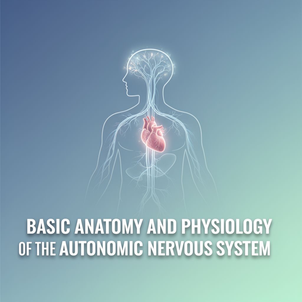
Understanding the structure and function of the human body's systems is foundational for optimizing athletic training and developing effective strength and conditioning programs. Physical exercise and sport performance rely on effective, purposeful movements generated by muscular forces acting through the skeletal system. This complex process is controlled by the brain's cerebral cortex, which activates skeletal muscle cells via motor neurons of the peripheral nervous system. Supporting this neuromuscular activity requires continuous delivery of oxygen and nutrients to working tissues and efficient removal of carbon dioxide and metabolic waste products, tasks handled by the cardiovascular and respiratory systems.
This guide will summarize the essential aspects of the musculoskeletal, neuromuscular, cardiovascular, and respiratory systems, which are critical for developing and maintaining muscular force and power.
🦴 Musculoskeletal System
The musculoskeletal system enables movement and provides structural support for the body.
1️⃣ Components and Function
The human musculoskeletal system comprises bones, joints, muscles, and tendons, configured to allow a wide variety of movements.
- Muscles generate force by pulling on bones, which act as levers around joints. Muscles can only pull, not push.
- Bones provide leverage, support, and protection.
2️⃣ Skeleton
The human body has approximately 206 bones, categorized into two main divisions:
- Axial Skeleton 🦴:
- Consists of the skull (cranium), vertebral column (C1 to coccyx), ribs, and sternum.
- Appendicular Skeleton 🦵:
- Includes the shoulder (pectoral) girdle (scapula, clavicle), bones of the arms, wrists, and hands (humerus, radius, ulna, carpals, metacarpals, phalanges).
- Pelvic girdle (coxal/innominate bones).
- Bones of the legs, ankles, and feet (femur, patella, tibia, fibula, tarsals, metatarsals, phalanges).
3️⃣ Joints
Joints are junctions between bones, classified by their degree of movement:
- Fibrous Joints (e.g., skull sutures) 🚫: Allow virtually no movement.
- Cartilaginous Joints (e.g., intervertebral disks) ↔️: Allow limited movement.
- Synovial Joints (e.g., elbow, knee, shoulder) ✅: Allow considerable movement.
- Most sport and exercise movements occur at synovial joints.
- Key features: Low friction and large range of motion.
- Articulating bone ends are covered with smooth hyaline cartilage.
- The entire joint is enclosed in a capsule filled with synovial fluid.
- Often supported by ligaments and additional cartilage.
Joint Movement & Axes 🔄
Joints are categorized by the number of directions (axes) about which rotation can occur:
- Uniaxial Joints (e.g., elbow): Operate as hinges, rotating about one axis.
- Biaxial Joints (e.g., ankle, wrist): Allow movement about two perpendicular axes.
- Multiaxial Joints (e.g., shoulder, hip ball-and-socket): Allow movement about all three perpendicular axes.
Vertebral Column
Composed of vertebral bones separated by flexible disks, allowing movement.
- Cervical vertebrae (7): Neck region.
- Thoracic vertebrae (12): Middle to upper back.
- Lumbar vertebrae (5): Lower back.
- Sacral vertebrae (5, fused): Rear part of the pelvis.
- Coccygeal vertebrae (3-5): Vestigial internal tail.
4️⃣ Factors Affecting Skeletal Growth in Adults 📈
Bone density and mineral content can be positively affected by:
- Heavy loads: From job tasks or resistance training.
- Explosive movements with impact: Activities like gymnastics with hard landings.
- Axial skeleton loading: How often and intensely the spine and pelvis are loaded.
- Varying stimulus: Bone adaptation is slower than muscle, so frequency, intensity, and type of loading should be varied.
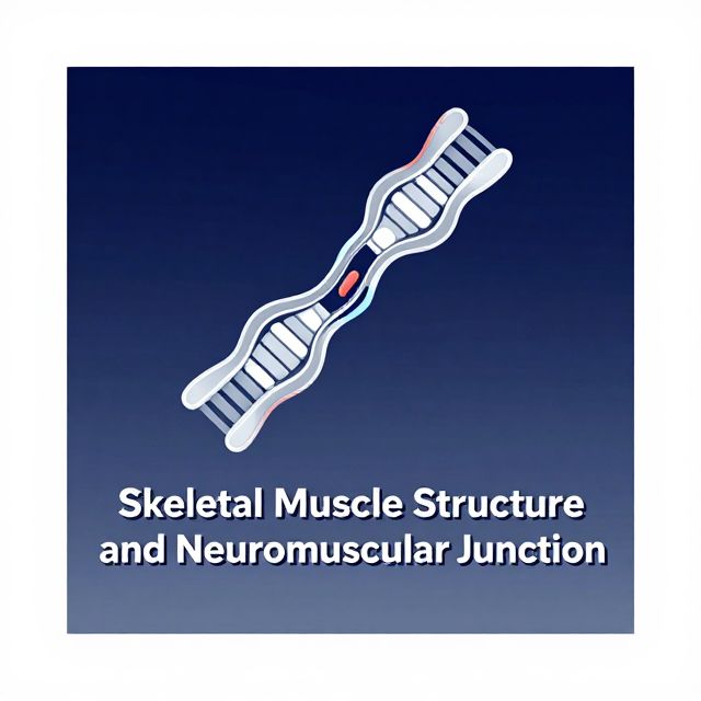
💪 Skeletal Musculature and Neuromuscular Control
This section details the structure of skeletal muscles and how the nervous system controls their contraction.
1️⃣ Musculoskeletal Macrostructure
Each skeletal muscle is an organ containing muscle tissue, connective tissue, nerves, and blood vessels.
- Epimysium (fibrous connective tissue): Covers the entire muscle.
- Tendon: Contiguous with the epimysium, attaches muscle to bone.
- Bone Periosteum: Specialized connective tissue covering all bones, where tendons attach. Muscle contraction pulls on the tendon, which then pulls on the bone.
- Attachments:
- Limb muscles: Proximal (closer to trunk) and Distal (farther from trunk).
- Trunk muscles: Superior (closer to head) and Inferior (closer to feet).
2️⃣ Musculoskeletal Microstructure
- Muscle Fibers (Muscle Cells): Long, cylindrical cells (50-100 µm diameter) with multiple nuclei, appearing striated.
- Fasciculi: Bundles of up to 150 muscle fibers, surrounded by perimysium.
- Endomysium: Connective tissue surrounding individual muscle fibers, contiguous with the sarcolemma (fiber's membrane).
- Connective Tissue Continuity: Epimysium, perimysium, and endomysium are all continuous with the tendon, transmitting tension from muscle cells to bone.
3️⃣ Neuromuscular Junction (Motor End Plate) 🧠
- The connection point between a motor neuron (nerve cell) and the muscle fibers it innervates.
- Each muscle cell has one neuromuscular junction.
- A single motor neuron can innervate many muscle fibers (hundreds to thousands).
- Motor Unit 📚: A motor neuron and all the muscle fibers it innervates. All fibers in a motor unit contract together when stimulated.
4️⃣ Inside the Muscle Fiber
- Sarcoplasm: Cytoplasm of a muscle fiber, containing contractile components, glycogen, fat, enzymes, mitochondria, and sarcoplasmic reticulum.
- Myofibrils: Hundreds dominate the sarcoplasm, containing the contractile apparatus.
- Myofilaments:
- Myosin (thick filaments): Contain up to 200 myosin molecules. Globular heads protrude, forming crossbridges that interact with actin.
- Actin (thin filaments): Two strands arranged in a double helix.
- Sarcomere 📚: The smallest contractile unit of skeletal muscle, where myosin and actin filaments are organized longitudinally.
- M-bridge: Anchors adjacent myosin filaments in the center.
- Z-line: Anchors actin filaments at both ends of the sarcomere.
- A-band: Dark band, corresponds to the alignment of myosin filaments.
- I-band: Light band, contains only actin filaments. Z-line is in the middle.
- H-zone: Area in the center of the sarcomere where only myosin filaments are present.
- Sarcoplasmic Reticulum (SR): Intricate system of tubules surrounding each myofibril, storing calcium ions (Ca²⁺).
- T-tubules (Transverse Tubules): Run perpendicular to the SR, transmitting action potentials from the sarcolemma deep into the muscle fiber, triggering Ca²⁺ release.
5️⃣ Sliding-Filament Theory of Muscular Contraction 💡
This theory explains how muscle fibers shorten.
- Mechanism: Actin filaments at each end of the sarcomere slide inward over myosin filaments, pulling the Z-lines toward the center of the sarcomere, thus shortening the muscle fiber.
- Crossbridge Action: Myosin crossbridges pull on the actin filaments. Rapid, repeated flexions of many crossbridges cause measurable movement.
- H-zone and I-band: Both shrink during contraction.
- Force Production: The amount of force produced is directly related to the number of myosin crossbridges bound to actin filaments at any given instant.
Phases of Muscle Contraction 1️⃣2️⃣3️⃣
- Resting Phase: Little calcium in myofibril (stored in SR). Myosin and actin form a weak bond.
- Excitation-Contraction Coupling Phase:
- Action potential signals Ca²⁺ release from SR.
- Ca²⁺ binds to troponin (protein on actin filament).
- This causes a shift in tropomyosin (protein running along actin), exposing actin binding sites.
- Myosin crossbridges rapidly attach to actin.
- Contraction Phase (Power Stroke):
- Energy from ATP hydrolysis (ATP → ADP + Pᵢ), catalyzed by myosin ATPase.
- Myosin head changes shape, pulling the actin filament toward the sarcomere center.
- ADP is released.
- Recharge Phase:
- Another ATP molecule binds to the myosin head, causing it to detach from actin.
- Myosin head resets to its original position.
- This cycle (binding, power stroke, dissociation, resetting) repeats as long as Ca²⁺ and ATP are available.
- Relaxation Phase:
- Motor nerve stimulation stops.
- Ca²⁺ is pumped back into the SR.
- Troponin and tropomyosin return to their resting positions, blocking actin binding sites.
- Actin and myosin filaments return to their unbound state.
- ⚠️ Key Point: Calcium and ATP are essential for crossbridge cycling.
6️⃣ Activation of Muscles
- All-or-None Principle 📚: When a motor neuron fires an impulse, all muscle fibers in its motor unit are simultaneously activated and develop force. A stronger action potential does not produce a stronger contraction.
- Twitch: A brief contraction resulting from a single action potential.
- Summation: If a second twitch is elicited before complete relaxation, forces summate, producing greater force.
- Tetanus 📚: High-frequency stimulation causes twitches to merge and completely fuse, representing the maximal force a motor unit can develop.
7️⃣ Muscle Fiber Types 📊
Skeletal muscles contain fibers with distinct morphological and physiological characteristics.
- Classification:
- Type I (Slow-Twitch): Develop force and relax slowly, long twitch time.
- Type IIa (Fast-Twitch): Rapid force development and relaxation, short twitch time.
- Type IIx (Fast-Twitch): Even faster and more powerful than Type IIa.
- Motor Unit Composition: A motor unit is composed of muscle fibers of the same type.
Major Characteristics of Muscle Fiber Types (Table 1.1 Summary)
| Characteristic | Type I (Slow-Twitch) | Type IIa (Fast-Twitch) | Type IIx (Fast-Twitch) | | :------------------------- | :------------------- | :--------------------- | :--------------------- | | Motor Neuron Size | Small | Large | Large | | Recruitment Threshold | Low | Intermediate/High | High | | Nerve Conduction Velocity | Slow | Fast | Fast | | Contraction Speed | Slow | Fast | Fast | | Relaxation Speed | Slow | Fast | Fast | | Fatigue Resistance | High | Intermediate/Low | Low | | Endurance | High | Intermediate/Low | Low | | Force Production | Low | Intermediate | High | | Power Output | Low | Intermediate/High | High | | Aerobic Enzyme Content | High | Intermediate/Low | Low | | Anaerobic Enzyme Content | Low | High | High | | Sarcoplasmic Reticulum Complexity | Low | Intermediate/High | High | | Capillary Density | High | Intermediate | Low | | Myoglobin Content | High | Low | Low | | Mitochondrial Size, Density | High | Intermediate | Low | | Fiber Diameter | Small | Intermediate | Large | | Color | Red | White/Red | White |
- 💡 Insight: Postural muscles (e.g., soleus) have high Type I composition. Locomotor muscles (e.g., quadriceps) have a mix for varied power output.
8️⃣ Motor Unit Recruitment Patterns
Muscular force can be graded in two ways:
- Variation in Activation Frequency: Increasing the frequency of motor unit activation leads to summation and greater force. Important in small muscles.
- Recruitment of Additional Motor Units: Increasing the number of activated motor units. Important in large muscles.
- Henneman's Size Principle (implied): Motor units are recruited in an orderly fashion, from smallest (Type I) to largest (Type IIx), depending on the force requirement.
- Activity-Specific Recruitment: Slow-twitch units for endurance, fast-twitch for high force/power.
- Untrained individuals may not fully activate the available motor neuron pool for maximal force.
Relative Involvement of Muscle Fiber Types in Sport Events (Table 1.2 Examples)
| Event | Type I | Type II | | :-------------------------- | :----- | :------ | | 100 m sprint | Low | High | | 800 m run | High | High | | Marathon | High | Low | | Olympic weightlifting | Low | High | | Soccer, lacrosse, hockey | High | High | | American football wide receiver | Low | High | | American football lineman | Low | High | | Basketball, team handball | Low | High | | Volleyball | Low | High | | Baseball or softball pitcher | Low | High | | Boxing | High | High | | Wrestling | High | High | | 50 m swim | Low | High | | Field events | Low | High | | Cross-country skiing, biathlon | High | Low | | Tennis | High | High | | Downhill or slalom skiing | High | High | | Speed skating | High | High | | Track cycling | Low | High | | Distance cycling | High | Low | | Rowing | High | High |
9️⃣ Proprioception proprioceptors
📚 Proprioceptors: Specialized sensory receptors located within joints, muscles, and tendons that provide the central nervous system (CNS) with information about body position, movement, and muscle tension. This information is crucial for maintaining muscle tone and coordinating complex movements.
-
Muscle Spindles 🦵:
- Location: Within muscle belly, parallel to normal (extrafusal) muscle fibers.
- Structure: Consist of modified muscle fibers (intrafusal fibers) enclosed in a connective tissue sheath.
- Function: Detect muscle length and rate of change in length. When a muscle lengthens, spindles stretch, activating sensory neurons that send impulses to the spinal cord, leading to reflexive muscle activation (e.g., knee jerk reflex).
- Role: Facilitate muscle activation; greater stretch leads to greater activation. Muscles performing precise movements have more spindles.
-
Golgi Tendon Organs (GTOs) 🔗:
- Location: In tendons, near the myotendinous junction, in series with extrafusal muscle fibers.
- Function: Detect tension in the tendon caused by muscle contraction. As tension increases, GTOs discharge, activating an inhibitory interneuron in the spinal cord. This inhibits the motor neuron serving the same muscle, causing the muscle to relax.
- Role: Protective mechanism against excessive tension. At low forces, their effect is minimal, but under extremely heavy loads, they cause muscle relaxation. Heavy resistance training can help the motor cortex override this inhibition.
🔟 How Athletes Can Improve Force Production 🚀
- Optimize Neural Recruitment: Incorporate phases of training with heavier loads.
- Increase Muscle Cross-Sectional Area: Build muscle mass.
- Recruit Fast-Twitch Fibers: Perform multi-muscle, multi-joint exercises with explosive actions.
❤️ Cardiovascular System
The cardiovascular system is vital for transporting substances throughout the body and maintaining internal homeostasis.
1️⃣ Primary Roles 🩸
- Transport nutrients and remove waste products.
- Assist with maintaining the body's internal environment.
- Regulate acid-base balance, fluids, and temperature.
2️⃣ Heart
A muscular organ composed of two interconnected but separate pumps:
- Right Side: Pumps deoxygenated blood through the lungs (pulmonary circulation).
- Left Side: Pumps oxygenated blood through the rest of the body (peripheral circulation).
Heart Chambers
- Atria (Right & Left): Receive blood and deliver it to the ventricles.
- Ventricles (Right & Left): Supply the main force for moving blood.
Heart Valves 🚪
Prevent backflow of blood. They open and close passively based on pressure gradients.
- Atrioventricular (AV) Valves:
- Tricuspid Valve: Between right atrium and right ventricle.
- Mitral (Bicuspid) Valve: Between left atrium and left ventricle.
- Prevent backflow into atria during ventricular contraction (systole).
- Semilunar Valves:
- Aortic Valve: Between left ventricle and aorta.
- Pulmonary Valve: Between right ventricle and pulmonary artery.
- Prevent backflow from arteries into ventricles during ventricular relaxation (diastole).
3️⃣ Conduction System ⚡
A specialized electrical system controls the heart's mechanical contraction.
- Sinoatrial (SA) Node 📚: The intrinsic pacemaker, normally initiating rhythmic electrical impulses (60-80 bpm).
- Internodal Pathways: Conduct impulses from SA node to AV node.
- Atrioventricular (AV) Node: Delays the impulse slightly, allowing atria to contract before ventricles (40-60 bpm).
- Atrioventricular (AV) Bundle (Bundle of His): Conducts impulse to ventricles.
- Left & Right Bundle Branches: Further divide into Purkinje Fibers, rapidly conducting impulses to all parts of the ventricles, causing simultaneous contraction.
Heart Rate Regulation
- Autonomic Nervous System: Influences myocardium rhythmicity.
- Sympathetic Nervous System: Accelerates SA node depolarization, increasing heart rate.
- Parasympathetic Nervous System: Slows SA node discharge, decreasing heart rate.
- Normal Resting Heart Rate: 60-100 beats/min.
- Bradycardia 📚: < 60 beats/min.
- Tachycardia 📚: > 100 beats/min.
4️⃣ Electrocardiogram (ECG) 📊
A graphic representation of the heart's electrical activity.
- P-wave: Electrical depolarization of the atria, leading to atrial contraction.
- QRS Complex: Electrical depolarization of the ventricles, leading to ventricular contraction. (Atrial repolarization is masked here).
- T-wave: Electrical repolarization of the ventricles (recovery from depolarization).
5️⃣ Blood Vessels
A single closed-circuit system with two components:
- Arterial System: Carries blood away from the heart.
- Arteries: Rapidly transport high-pressure blood from the heart; strong, muscular walls.
- Arterioles: Small branches of arteries, act as control vessels, regulating blood flow to capillaries. Strong, muscular walls allow significant constriction/dilation.
- Venous System: Returns blood toward the heart.
- Capillaries: Facilitate exchange of oxygen, nutrients, waste, etc., between blood and interstitial fluid. Very thin, permeable walls.
- Venules: Collect blood from capillaries.
- Veins: Transport blood back to the heart; thin, muscular walls. Low pressure, can constrict/dilate to act as a blood reservoir. Many veins (especially in legs) have one-way valves to prevent retrograde blood flow.
Skeletal Muscle Pump 💡
Contracting muscles assist the venous system in returning blood to the heart. Muscle contraction compresses veins, and one-way valves ensure blood flows only towards the heart. This prevents blood pooling in lower extremities, especially after exercise or during prolonged sitting.
6️⃣ Blood
- Primary Functions:
- Transport oxygen from lungs to tissues for cellular metabolism (via hemoglobin in red blood cells).
- Remove carbon dioxide (most abundant metabolic by-product) from tissues to lungs.
- Hemoglobin: Iron-protein molecule, also acts as an acid-base buffer (regulates hydrogen ion concentration).
- Red Blood Cells: Contain carbonic anhydrase, which catalyzes CO₂ and water reaction to facilitate CO₂ removal.
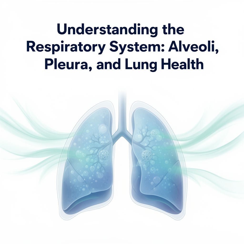
🌬️ Respiratory System
The respiratory system is responsible for gas exchange between the body and the external environment.
1️⃣ Primary Function 💨
- Basic exchange of oxygen (O₂) and carbon dioxide (CO₂).
2️⃣ Anatomy of Airway
Air passes through:
- Nose: Warms, humidifies, and purifies air.
- Pharynx
- Larynx
- Trachea (first-generation passage)
- Bronchi (right and left main bronchi are second-generation passages)
- Bronchioles (subsequent generations, approximately 23 divisions)
- Alveoli 📚: Tiny air sacs where gas exchange occurs.
3️⃣ Exchange of Air (Ventilation)
The amount and movement of air in and out of the lungs.
- Lung Expansion/Recoil: Lungs do not actively expand; they are acted upon by:
- Diaphragm Movement: Downward (inspiration) and upward (expiration) movement lengthens/shortens the chest cavity. Normal, quiet breathing is primarily diaphragmatic.
- Rib Cage Elevation/Depression: Increases/decreases the front-to-back diameter of the chest cavity.
- Muscles of Inspiration: Elevate the rib cage (external intercostals, sternocleidomastoids, anterior serrati, scaleni).
- Muscles of Expiration: Depress the chest (abdominal muscles: rectus abdominis, obliques, transversus abdominis; internal intercostals). During heavy breathing, abdominal muscles push the diaphragm up.
- Pleural Pressure: Pressure in the space between lung pleura and chest wall pleura. Normally slightly negative. Becomes more negative during inspiration, pulling on lungs.
- Alveolar Pressure: Pressure inside the alveoli. Must fall slightly below atmospheric pressure for inspiration, and rise above for expiration.
- Energy Expenditure: Normal breathing uses 3-5% of total energy; heavy exercise can increase this to 8-15%, especially with airway resistance (e.g., exercise-induced asthma).
Training Respiratory Muscles 🏋️♀️
- Regular exercise (endurance, resistance) generally maintains respiratory muscle function.
- Resistance exercise, especially with Valsalva maneuver, taxes the diaphragm and abdominal muscles.
- Specific respiratory muscle training is usually not necessary unless recovering from surgery, prolonged bed rest, or compromised breathing patterns.
4️⃣ Exchange of Respiratory Gases (Diffusion)
- Mechanism: Gases move by simple random motion from a region of high concentration (or partial pressure) to a region of low concentration.
- Oxygen Diffusion: O₂ diffuses from alveoli (high partial pressure) into pulmonary blood (low partial pressure).
- Carbon Dioxide Diffusion: CO₂ diffuses from blood (high partial pressure) into alveoli (low partial pressure).
- This process is extremely rapid.
✅ Conclusion: Importance for Strength and Conditioning Professionals
A thorough understanding of the anatomy and physiology of the musculoskeletal, neuromuscular, cardiovascular, and respiratory systems is paramount for strength and conditioning professionals. This knowledge forms the scientific basis for designing effective training programs. It includes comprehending:
- The function of macro- and microstructure of the skeleton and muscle fibers.
- Different muscle fiber types and their roles.
- Interactions between tendons and muscles.
- The motor unit and its activation patterns.
- The integrated functions of the heart, vascular system, lungs, and respiratory mechanics.
This comprehensive understanding allows professionals to develop training strategies tailored to an athlete's specific needs, maximizing performance and minimizing injury risk.
📚 Key Terms
- A-band
- Acetylcholine
- Actin
- Action potential
- All-or-none principle
- Alveolar pressure
- Alveoli
- Aortic valve
- Appendicular skeleton
- Arterial system
- Arteriole
- Artery
- Atrioventricular (AV) bundle
- Atrioventricular (AV) node
- Atrioventricular (AV) valves
- Atrium
- Axial skeleton
- Biaxial joints
- Bone periosteum
- Bradycardia
- Bronchi
- Bronchiole
- Capillary
- Cartilaginous joints
- Crossbridge
- Depolarization
- Diastole
- Diffusion
- Distal
- Electrocardiogram (ECG)
- Endomysium
- Epimysium
- Extrafusal fibers
- Fasciculi
- Fast-twitch fiber
- Fibrous joints
- Golgi tendon organ (GTO)
- Hemoglobin
- Hyaline cartilage
- H-zone
- I-band
- Inferior
- Intrafusal fibers
- Left bundle branch
- Mitral valve
- Motor neuron
- Motor unit
- Multiaxial joints
- Muscle fiber
- Muscle spindle
- Myocardium
- Myofibril
- Myofilament
- Myosin
- Neuromuscular junction
- Parasympathetic nervous system
- Perimysium
- Pleura
- Pleural pressure
- Power stroke
- Proprioceptor
- Proximal
- Pulmonary valve
- Purkinje fibers
- P-wave
- QRS complex
- Red blood cell
- Repolarization
- Right bundle branch
- Sarcolemma
- Sarcomere
- Sarcoplasm
- Sarcoplasmic reticulum
- Semilunar valves
- Sinoatrial (SA) node
- Sliding-filament theory
- Slow-twitch fiber
- Superior
- Sympathetic nervous system
- Synovial fluid
- Synovial joints
- Systole
- Tachycardia
- Tendon
- Tetanus
- Trachea
- Tricuspid valve
- Tropomyosin
- Troponin
- T-tubule
- T-wave
- Twitch
- Type I fiber
- Type IIa fiber
- Type IIx fiber
- Uniaxial joints
- Vein
- Venous system
- Ventricle
- Venule
- Vertebral column
- Z-line
📝 Study Questions
-
Which of the following substances regulates muscle actions? a. potassium b. calcium c. troponin d. tropomyosin
-
Which of the following substances acts at the neuromuscular junction to excite the muscle fibers of a motor unit? a. acetylcholine b. ATP c. creatine phosphate d. serotonin
-
When throwing a baseball, an athlete’s arm is rapidly stretched just before throwing the ball. Which of the following structures detects and responds to that stretch by reflexively increasing muscle activity? a. Golgi tendon organ b. muscle spindle c. extrafusal muscle d. Pacinian corpuscle
-
From which of the following is the heart’s electrical impulse normally initiated? a. AV node b. SA node c. the brain d. the sympathetic nervous system
-
Which of the following occurs during the QRS complex of a typical ECG? I. depolarization of the atrium II. repolarization of the atrium III. repolarization of the ventricle IV. depolarization of the ventricle a. I and III only b. II and IV only c. I, II, and III only d. II, III, and IV only