This study material has been compiled from various sources, including copy-pasted text and a lecture audio transcript, to provide a comprehensive overview of the oral region.
📚 Anatomy and Physiology of the Oral Cavity
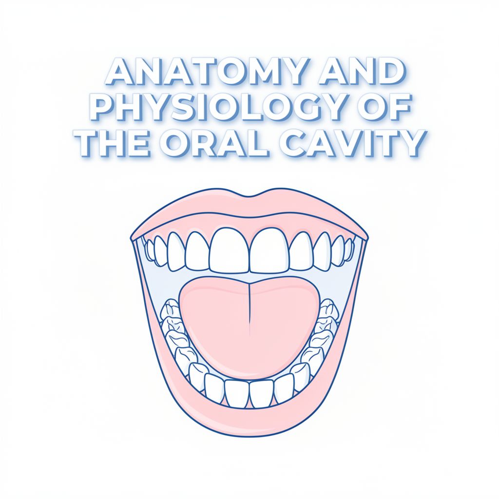
🎯 Introduction to the Oral Region
The oral region is a complex and critical area for initial digestion and various sensory functions. It encompasses the oral cavity itself, the teeth, gingivae (gums), tongue, palate, and the region of the palatine tonsils. This anatomical space is where food is first ingested and prepared for subsequent digestion in the stomach and small intestine.
✅ Key Processes in the Oral Cavity:
- Ingestion: Taking food into the body.
- Mastication (Chewing): Mechanical breakdown of food by teeth.
- Salivation: Saliva from salivary glands moistens food and forms a manageable food bolus.
- Deglutition (Swallowing): Voluntarily initiated process where the bolus is pushed from the oral cavity into the pharynx, transitioning to an involuntary phase.
The oral cavity, commonly known as the mouth, is structurally divided into two main parts:
- Oral Vestibule
- Oral Cavity Proper
👄 Components of the Oral Cavity
1. Oral Vestibule
The oral vestibule is a slit-like space located between the teeth and gingivae (gums) and the lips and cheeks.
- Communication: It communicates with the exterior through the oral fissure (mouth opening).
- Control: The size of the oral fissure is controlled by circumoral muscles:
- Orbicularis oris: Acts as the sphincter of the oral fissure.
- Buccinator, Risorius, Depressors and Elevators of the lips: Function as dilators of the fissure.
2. Oral Cavity Proper
The oral cavity proper is the space situated between the upper and lower dental arches (maxillary and mandibular alveolar arches and the teeth they bear).
- Boundaries: Limited laterally and anteriorly by the dental arches.
- Roof: Formed by the palate.
- Posterior Communication: Communicates with the oropharynx (oral part of the pharynx).
- Resting State: When the mouth is closed and at rest, the oral cavity proper is fully occupied by the tongue.
3. Lips
The lips are mobile, musculofibrous folds surrounding the mouth.
- Structure: Extend from the nasolabial sulci and nares laterally and superiorly to the mentolabial sulcus inferiorly. They contain the orbicularis oris, superior and inferior labial muscles, vessels, and nerves.
- Covering: Covered externally by skin and internally by mucous membrane.
- Functions:
- Act as valves of the oral fissure, controlling entry and exit.
- Grasping food, sucking liquids.
- Keeping food out of the vestibule.
- Forming speech.
- Osculation (kissing).
- Blood Supply:
- Superior and inferior labial arteries (branches of facial arteries) anastomose to form an arterial ring.
- Upper lip: Superior labial branches of facial and infra-orbital arteries.
- Lower lip: Inferior labial branches of facial and mental arteries.
- Venous Drainage: Superior and inferior labial veins drain into the facial vein, then into the common facial vein.
- Nerve Supply:
- Upper lip: Superior labial branches of the infra-orbital nerve (CN V2).
- Lower lip: Inferior labial branches of the mental nerve (CN V3).
- Lymphatic Drainage:
- Upper lip and lateral lower lip: Primarily to submandibular lymph nodes.
- Medial lower lip: Initially to submental lymph nodes.
4. Cheeks
The cheeks have a similar structure to the lips and form the movable walls of the oral cavity.
- Anatomical Region: The external aspect constitutes the buccal region.
- Boundaries:
- Anteriorly: Oral and mental regions (lips and chin).
- Superiorly: Zygomatic region.
- Posteriorly: Parotid region.
- Inferiorly: Inferior border of the mandible.
- Prominence: Occurs at the junction of the zygomatic and buccal regions, often referred to as the "cheek bone" (zygomatic bone and arch).
- Muscles: Principal muscles are the buccinators.
- Glands & Fat: Numerous small buccal glands lie between the mucous membrane and buccinators. Encapsulated buccal fat-pads are superficial to the buccinators, larger in infants to reinforce cheeks during sucking.
- Blood Supply: Buccal branches of the maxillary artery.
- Innervation: Buccal branches of the mandibular nerve.
5. Gingivae (Gums)
The gingivae are composed of fibrous tissue covered with mucous membrane.
- Gingiva Proper (Attached Gingiva): Firmly attached to the alveolar processes of the mandible and maxilla and the necks of the teeth.
- Adjacent to tongue: Superior and inferior lingual gingivae.
- Adjacent to lips/cheeks: Maxillary and mandibular labial or buccal gingiva.
- Characteristics: Normally pink, stippled, and keratinizing.
- Alveolar Mucosa (Unattached Gingiva): Normally shiny red and non-keratinizing.
- Blood Supply:
- Maxillary Artery Branches: Anterior/posterior superior alveolar arteries (upper gums), greater palatine artery (palate).
- Facial Artery Branches: Submental artery (lower lip, lingual gingiva).
- Lingual Artery Branches: Sublingual artery (floor of mouth, lower lingual gingiva).
- Inferior Alveolar Artery: Supplies lower jaw gingiva and teeth, with mental and incisive branches for the lower lip area.
- Venous Drainage: Gingival plexus (network of tiny blood vessels), veins (buccal, lingual) drain into pterygoid plexus or internal jugular vein.
- Nerve Supply:
- Lower Gums (Mandibular Division - V3): Lingual nerve (inner gingiva), Inferior Alveolar Nerve (lower teeth, periodontal ligaments, facial gingiva via mental nerve branch), Buccal nerve (outer gingiva).
- Upper Gums (Maxillary Division - V2): Superior Alveolar Nerves (gingiva around upper teeth).
🦷 The Teeth
1. General Information
Teeth are set in tooth sockets, primarily used for mastication and assisting in articulation (speech).
- Classification: Deciduous (primary) or Permanent (secondary).
- Identification: By type and proximity to the midline (e.g., incisors, 1st molar).
- Functions:
- Incise, reduce, and mix food with saliva during chewing.
- Help sustain themselves by supporting surrounding tissues.
- Participate in articulation.
- Number:
- Children: 20 deciduous teeth.
- Adults: 32 permanent teeth.
- Types by Characteristics:
- Incisors: Thin cutting edges.
- Canines: Single prominent cones.
- Premolars (Bicuspids): Two cusps.
- Molars: Three or more cusps.
2. Tooth Eruption
📚 Definition: The natural process where developing teeth move from the jawbone through the gums into the mouth, becoming visible and functional.
- Timeline:
- Baby teeth: Start around 6 months, all 20 by age 3.
- Permanent teeth: Begin around age 6 (first molars, lower incisors), continue through teens, all adult teeth (including wisdom teeth) typically by early twenties.
- Order: Lower front teeth (incisors) usually first, then upper front, molars, canines, and finally second molars. Permanent teeth generally follow a similar order, replacing baby teeth.
3. Parts and Structure of Teeth
Tooth anatomy includes the visible crown, embedded root, and connecting neck.
- Hard Tissues:
- Enamel: Outer layer of the crown, hardest substance in the body.
- Dentin: Surrounds the pulp, forms most of the tooth.
- Cementum: Covers the root, anchors it to the jawbone.
- Soft Tissue:
- Pulp: Contains nerves, blood vessels, and connective tissue.
- Supporting Structures:
- Periodontal Ligament: Connects cementum to the jawbone.
- Gingiva (Gums): Covers the supportive periodontium.
- Regions:
- Crown: Projects from the gingiva.
- Neck: Between the crown and root.
- Root: Fixed in the tooth socket by the periodontium; number varies.
- Internal Structures:
- Pulp Cavity: Contains connective tissue, blood vessels, and nerves.
- Root Canal (Pulp Canal): Transmits nerves and vessels to/from the pulp cavity.
- Apical Foramen: Opening at the root apex for nerve and vessel passage.
4. Tooth Sockets
The tooth sockets are in the alveolar processes of the maxillae and mandible.
- Septa: Adjacent sockets separated by interalveolar septa; roots of multi-rooted teeth separated by interradicular septa.
- Bone Thickness: Labial wall thin over incisors, lingual wall thin over molars.
- Clinical Implication: Labial surface commonly broken for incisor extraction, lingual for molar extraction.
5. Dento-alveolar Syndesmosis (Gomphosis) / Periodontium
📚 Definition: A special type of fibrous joint connecting tooth roots to the alveolar bone.
- Periodontium (Periodontal Membrane): Composed of collagenous fibers between the root's cement and the alveolus's periosteum.
- Rich Supply: Abundantly supplied with tactile, pressoreceptive nerve endings, lymph capillaries, and glomerular blood vessels.
- Function: Acts as hydraulic cushioning to curb axial masticatory pressure.
- Pressoreceptive Nerve Endings: Detect changes in pressure.
6. Vasculature of Teeth
The tooth's vasculature, mainly in the pulp, comes from the external carotid artery via the maxillary artery.
- Arterial Supply:
- Superior alveolar arteries (upper teeth).
- Inferior alveolar arteries (lower teeth).
- Enter through apical foramina, forming a rich capillary network in the pulp for nutrient supply and waste removal.
- Venous Drainage: Alveolar veins (same names as arteries) accompany the arteries.
- Lymphatic Drainage: From teeth and gingivae, mainly to submandibular lymph nodes.
7. Nerve Supply to Teeth
Primarily from the Trigeminal Nerve (CN V).
- Branches: Maxillary (V2) and Mandibular (V3) branches split into superior and inferior alveolar nerves.
- Plexuses: Form superior and inferior dental plexuses.
- Sensory Input: Deliver sensory input (pain, temperature, pressure) from pulp and surrounding tissues.
- Raschkow's Nerve Plexus: Sensory nerve fibers travel through the apical foramen into the pulp, forming this plexus, allowing for sharp pain and dull ache sensations.
👅 The Palate
The palate is the roof of the mouth, separating the oral cavity from the nasal cavity.
- Crucial for: Eating, breathing, swallowing, and speech.
- Surfaces: Superior (nasal) surface covered with respiratory mucosa; inferior (oral) surface covered with oral mucosa densely packed with glands.
1. Hard Palate
The anterior two-thirds of the palate has a bony skeleton.
- Structure: Vaulted (concave), mostly filled by the tongue at rest. Formed by palatine processes of the maxillae and horizontal plates of the palatine bones.
- Incisive Fossa: Depression posterior to central incisors where incisive canals open.
- Nasopalatine Nerves: Pass from the nose through incisive canals and foramina into the incisive fossa.
- Greater Palatine Foramen: Medial to the 3rd molar, where greater palatine vessels and nerve emerge.
- Lesser Palatine Foramina: Posterior to greater palatine foramen, transmit lesser palatine nerves and vessels to the soft palate.
2. Soft Palate (Velum)
The movable posterior third of the palate, suspended from the hard palate's posterior border.
- Structure: Flexible, muscular, no bony skeleton. Anterior aponeurotic part strengthened by palatine aponeurosis.
- Uvula: Conical process hanging from the curved free margin posteroinferiorly.
- Function during Swallowing:
- Tensed to allow tongue to press against it, squeezing food bolus.
- Elevated posteriorly and superiorly against the pharynx wall, preventing food from entering the nasal cavity.
- Connections: Continuous laterally with the pharynx wall. Joined to the tongue by palatoglossal arches and to the pharynx by palatopharyngeal arches.
- Taste Buds: A few located in the epithelium covering the oral surface.
🗣️ The Tongue
The tongue is a muscular organ in the mouth, crucial for manipulating food, taste, speech, and cleaning teeth.
1. General Anatomy
- Structure: Muscular hydrostat, richly supplied with nerves and blood vessels.
- Divisions:
- Oral Part (Anterior 2/3): Visible part.
- Pharyngeal Part (Posterior 1/3): Closest to the throat.
- Median Sulcus: Groove on the dorsal surface, separating left and right sides by the lingual septum.
- Terminal Sulcus: A "V"-shaped groove dividing the anterior and posterior parts.
- Foramen Cecum: Blind foramen at the apex of the terminal sulcus, remnant of the thyroglossal diverticulum.
- Connections (Posterior Part): Connected to the hyoid bone, epiglottis (glossoepiglottic folds), soft palate (glossopalatine arches), and pharynx.
2. Upper Surface (Dorsum)
- Median Sulcus: Divides the dorsum into symmetrical halves.
- Terminal Sulcus: Divides the tongue into posterior pharyngeal part and anterior oral part.
- Nerve Supply (Dorsum):
- Pharyngeal part: Glossopharyngeal nerve (CN IX).
- Oral part: Lingual nerve (CN V3) for somatosensory perception; Chorda tympani (CN VII) for taste perception.
3. Taste Buds
Clusters of taste receptor cells (gustatory cells) located around papillae on the tongue, soft palate, upper esophagus, cheek, and epiglottis.
- Detection: Five elements of taste perception: saltiness, sourness, bitterness, sweetness, and savoriness (umami).
- Mechanism: Food dissolved in saliva contacts taste receptors via taste pores. Information sent to the brain via CN VII, IX, and X.
4. Types of Lingual Papillae
Raised protrusions on the tongue surface; all except one contain taste buds.
- 🍄 Fungiform Papillae: Mushroom-shaped, mostly on dorsal surface and sides. Innervated by facial nerve.
- 🌿 Foliate Papillae: Ridges and grooves on lateral borders towards the posterior part. Innervated by facial nerve (anterior) and glossopharyngeal nerve (posterior).
- ⭕ Circumvallate Papillae: 10-14 papillae at the back of the oral part, arranged in a circular row in front of the sulcus terminalis. Associated with Von Ebner's glands. Innervated by glossopharyngeal nerve.
- 📏 Filiform Papillae: Most numerous type, but do not contain taste buds. Involved in mechanical abrasion due to keratinization.
5. Undersurface (Ventral Surface)
- Frenulum: Fold of mucous membrane tethering the tongue to the floor of the mouth.
- Sublingual Caruncles: Small prominences on either side of the frenulum where submandibular glands drain.
6. Tongue Muscles
Eight muscles classified as intrinsic or extrinsic.
Extrinsic Muscles
Change the position of the tongue, anchored to bone.
- Genioglossus: Fan-shaped, forms majority of tongue body.
- Origin: Mental spine of mandible, some fibers from hyoid bone.
- Insertion: Hyoid bone, bottom of tongue.
- Innervation: Hypoglossal nerve (CN XII).
- Function: Protrudes tongue, deviates it to opposite side; depresses center of tongue when acting together.
- Hyoglossus:
- Origin: Hyoid.
- Insertion: Side of the tongue.
- Innervation: Hypoglossal nerve (CN XII).
- Actions: Depresses and retracts the tongue.
- Styloglossus:
- Origin: Styloid process of temporal bone.
- Insertion: Tip and sides of tongue.
- Innervation: Hypoglossal nerve (CN XII).
- Actions: Retraction and elevation of tongue.
- Palatoglossus:
- Origin: Palatine aponeurosis.
- Insertion: Tongue.
- Innervation: Vagus nerve (CN X) via pharyngeal branch to pharyngeal plexus.
- Actions: Raises the back part of the tongue.
Intrinsic Muscles
Change the shape of the tongue, originate and insert within the tongue itself.
- Superior Longitudinal Muscle: Shortens and curls the tongue upward, elevating tip and sides.
- Inferior Longitudinal Muscle: Shortens and curls the tongue downward, depressing tip and sides.
- Transverse Muscle: Narrows and elongates the tongue.
- Vertical Muscle: Flattens and widens the tongue. 💡 These muscles work together for flexibility, crucial for speech, mastication, and swallowing.
7. Blood Supply (Tongue)
Primary supply from the lingual artery (branch of external carotid artery), with contribution from the facial artery's tonsillar branch.
- Lingual Artery Branches:
- Dorsal Lingual Arteries: Supply posterior tongue, tonsil, soft palate, epiglottis.
- Sublingual Artery: Supplies intrinsic muscles and floor of the mouth.
8. Venous Drainage (Tongue)
- Dorsal Lingual Veins (from top/sides).
- Deep Lingual Veins (from tip/body).
- Form the Lingual Vein, emptying into the Internal Jugular Vein (IJV), often via the Sublingual Vein or Vena Comitans Nervi Hypoglossi.
9. Nerve Supply (Tongue)
The tongue's nerve supply is complex:
- Motor:
- Hypoglossal nerve (CN XII): Controls all tongue muscles, except palatoglossus.
- Vagus nerve (CN X): Innervates palatoglossus.
- Sensory:
- Lingual nerve (CN V3): General sensation for anterior 2/3.
- Facial nerve (CN VII) via chorda tympani: Taste for anterior 2/3.
- Glossopharyngeal nerve (CN IX): General sensation and taste for posterior 1/3.
- Superior Laryngeal nerve (CN X): Sensory input for a small part near the epiglottis.
🩺 Clinical Correlates of the Tongue
The tongue's appearance, function, and structure can reflect systemic health.
- Appearance & Color:
- Pale: Anemia, nutrient deficiency, coldness.
- Red/Bright Red: Infection, inflammation, excess internal heat, anxiety, insomnia.
- Bluish Tint: Poor circulation, blood congestion, coldness.
- Coating:
- White Coating: Fungal infections (thrush), dehydration, systemic issues.
- Hairy: Fungal overgrowth, certain medications, smoking.
- Texture/Shape:
- Cracked/Fissured: Dehydration, geographic tongue (benign), systemic disease.
- Geographic Tongue: Benign condition with irregular, red patches resembling a map.
- Size/Position: Can link to sleep apnea or developmental problems (e.g., "tongue-tie").
- Function: Tongue strength is linked to swallowing, speech, and even posture.