Study Material: Basic Chromosome Abnormalities, Hereditary Metabolic Diseases, and Cancer
Source Information: This study material has been compiled and organized from a lecture audio transcript and copy-pasted text content.
Introduction to Genetic Disorders 📚
Genetic disorders fundamentally arise from abnormalities in the number and structures of chromosomes. These abnormalities can be broadly categorized and examined in different groups, stemming primarily from errors during cell division. Understanding these basic concepts is crucial for comprehending human health and disease.
I. Chromosome Abnormalities 🧬
Genetic disorders often originate from issues with chromosomes, which can be numerical (changes in the total count) or structural (changes in their physical arrangement).
A. Numerical Abnormalities
These involve an incorrect number of chromosomes in a cell. The most significant mechanisms leading to numerical chromosome abnormalities are:
-
Non-disjunction:
- Mechanism: During cell division (specifically meiosis), chromosome pairs or sister chromatids fail to separate properly.
- Result: This leads to gametes (sperm or egg cells) having either an excess or a deficiency of chromosomes.
- Example: If a chromosome pair fails to separate in meiosis II, one gamete receives two chromosomes, and another receives none.
- Trisomy: When a gamete with two chromosomes unites with a normal gamete, the resulting zygote has three copies of a chromosome instead of the usual two.
- ✅ Examples: Down Syndrome (Trisomy 21), Klinefelter Syndrome (XXY).
- Monosomy: When a gamete lacking a chromosome unites with a normal gamete, the resulting zygote has only one copy of a chromosome.
- ✅ Examples: Turner Syndrome (XO), Monosomy G.
- Trisomy: When a gamete with two chromosomes unites with a normal gamete, the resulting zygote has three copies of a chromosome instead of the usual two.
-
Anaphase Lag:
- Mechanism: A chromosome or chromatid moves too slowly during anaphase and is consequently lost from the forming daughter nucleus.
- Result: This can also lead to trisomic or monosomic zygotes.
-
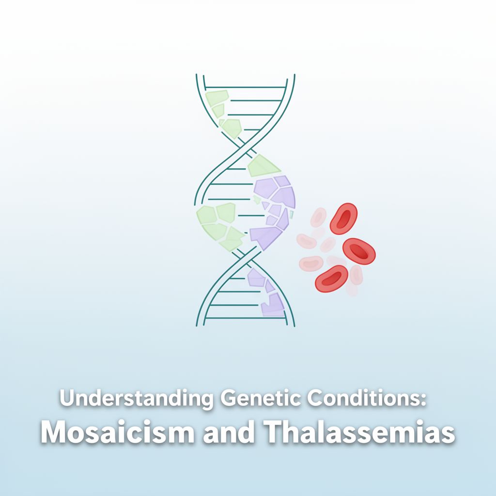
Mosaicism:
- Definition: A condition where different cells within the same individual have different numbers or arrangements of chromosomes. It's like a mosaic artwork where different tiles form a complete picture.
- Example: In mosaic trisomy 18, some cells have three copies of chromosome 18, while others have two.
-
Types of Ploidy:
- Euploidy: The number of chromosomes is an exact multiple of the normal haploid number (e.g., triploidy, tetraploidy). In humans, the haploid number is 23, so any addition beyond the diploid 46 chromosomes (like 69 or 92) is polyploidy.
- Aneuploidy: Involves the gain or loss of one or more individual chromosomes, not an entire set.
- Hypoploidy: Missing one or more chromosomes.
- Hyperploidy: Having one or more extra chromosomes.
B. Specific Syndromes Due to Numerical Abnormalities
-
Down Syndrome (Trisomy 21) 👶
- Cause: Approximately 95% of cases involve an additional 21st chromosome.
- Incidence: 1 in 700 live births, increasing significantly with maternal age (e.g., 1/2000 at early childbearing age vs. 45/1000 for mothers over 40).
- Paternal Contribution: In about one-third of cases, the extra 21st chromosome comes from the father.
- Symptoms: Extremely calm, rarely cries, hypotonicity (decreased muscle resistance), delayed physical and mental development (average IQ around 50).
-
Klinefelter Syndrome (XXY) ♂️
- Cause: Presence of an extra X chromosome in males (XXY genotype).
- Symptoms: Taller than average, infertile (unable to father biological children) due to small testes producing reduced testosterone.
- Associated Issues: Delayed/incomplete puberty, breast enlargement (gynecomastia), decreased muscle mass, decreased bone density, reduced facial/body hair.
- Health Risks: Increased risk of metabolic syndrome (type 2 diabetes, hypertension, increased belly fat), tremors, breast cancer (if gynecomastia develops), osteoporosis, and autoimmune disorders.
- Variations: More severe mental retardation and malformations occur with increasing numbers of X chromosomes (e.g., XXXY, XXXXY).
-
Turner Syndrome (XO) ♀️
- Cause: Affects females, characterized by the absence of all or part of one X chromosome (XO genotype).
- Symptoms: Short stature (evident by age 5), early loss of ovarian function (ovarian hypofunction or premature ovarian failure) leading to infertility in most cases.
- Note: A small percentage of affected females may retain normal ovarian function into young adulthood.
C. Structural Abnormalities of Chromosomes
These involve changes in the structure of one or more chromosomes.
- Translocation: A segment from one chromosome is transferred to a non-homologous chromosome or a new site on the same chromosome.
- Inversion: A chromosome segment is clipped out, turned upside down, and reinserted.
- Balanced: All genes are present; usually causes no problems.
- Unbalanced: Genes are deleted or duplicated; often associated with developmental delay, mental retardation, and birth defects.
- Duplication: Part of a chromosome is copied, resulting in one or more extra copies of a DNA segment, gene, or even an entire chromosome.
- Deletion: Loss of parts of chromosomes.
- ⚠️ Can cause severe congenital abnormalities and significant intellectual and physical disability.
D. Specific Deletion Syndromes
-
Wolf-Hirschhorn Syndrome (4p-)
- Cause: Deletion of genetic material near the end of the short (p) arm of chromosome 4.
- Severity: Varies with deletion size; larger deletions lead to more severe intellectual disability and physical abnormalities.
- Symptoms: Severe growth retardation, mental disorders, microcephaly, distinctive "Greek helmet" facial appearance, cleft lip and palate, coloboma (missing eye tissue), and cardiac septal defects.
-
Jacobsen Syndrome (11q deletion disorder)
- Cause: Deletion on the long arm of chromosome 11.
- Symptoms: Intellectual deficiency, distinct facial characteristics, cardiac defects, and physical problems including bleeding disorders.
-
Isochromosome: Forms when the centromere divides transversely instead of longitudinally, resulting in a chromosome with two identical arms (e.g., two long arms or two short arms).
-
Ring Chromosome: The two ends of a chromosome fuse together, forming a ring shape.
-
Gap: A visible break or distortion in the chromosome structure.
II. Hereditary Metabolic Diseases 🧪
These diseases arise from errors in genes at the molecular level, affecting the structure and function of proteins (often enzymes).
- ✅ Characteristics:
- Usually seen in the first days or weeks of life.
- Hereditary, often observed in cases of consanguineous marriage.
- Can range from mild to severe, impacting patients' lives to varying extents.
Examples of Metabolic Diseases:
-
Sickle Cell Anemia:
- Cause: Abnormal hemoglobin causes red blood cells to take on a sickle (semilunar) shape under low oxygen pressure.
- Symptoms: Anemic appearance, growth retardation in children.
-
Phenylketonuria (PKU) 🧠
- Cause: Inability to metabolize the amino acid phenylalanine due to a deficiency in the enzyme phenylalanine hydroxylase.
- Inheritance: Autosomal recessive. If both parents are carriers, there is a 25% chance their child will have PKU.
- Symptoms: Accumulation of phenylalanine damages the developing brain, leading to severe mental retardation and nervous system issues. Babies appear normal initially, but symptoms become evident around 5-6 months (e.g., delayed sitting, walking, talking; microcephaly).
- Diagnosis & Treatment: Early diagnosis via newborn screening is crucial. Treatment involves a strict diet low in phenylalanine, especially during the first 8-10 years of life when brain development is fastest, to prevent mental retardation.
-
Alkaptonuria:
- Cause: Deficiency of homogentisic acid oxidase, leading to the accumulation of homogentisic acid in connective tissues.
- Symptoms: Darkening of urine, pigmentation in cartilage, and development of arthritis resembling rheumatoid arthritis (but radiologically similar to osteoarthritis).
-
Albinism:
- Cause: Lack of the tyrosinase enzyme, preventing the synthesis of melanin pigment.
- Symptoms: Skin and hair lack pigment (white), extreme sensitivity to light due to lack of eye pigments.
III. Cancer 🦀
Cancer is a global health problem characterized by the uncontrolled growth and spread of abnormal cells.
- Definition (WHO): "One defining feature of cancer is the rapid creation of abnormal cells that grow beyond their usual boundaries, and which can then invade adjoining parts of the body and spread to other organs."
- Cause: Division of cells without complete maturation, due to hereditary changes at the cellular level.
- Treatment: Surgery and chemotherapy are common. Early diagnosis significantly improves outcomes.
- Prevention: Avoiding common risk factors like cigarette smoke.
A. Cellular Properties of Cancer Cells 📈
Cancerous cells acquire several new properties that they can pass on to their daughter cells:
- Loss of Division Control: They lose the ability to control their division and ignore inhibition mechanisms.
- Loss of Tissue Specificity: They lose their original tissue-specific properties and can spread to other tissues via blood or lymph (metastasis).
- Recessed Differentiation: They often manifest embryonal cell properties.
- Altered Metabolism: Increased sugar intake and aerobic respiration (known as the Warburg Effect – discovered by Otto Warburg in 1921, where tumor cells use extensive glucose and produce lactate even aerobically).
- Changed Antigenic Properties: Their surface antigens are altered.
- Changed Morphology: Their physical appearance is different from normal cells.
- Genomic Instability: Prone to further genetic mutations.
- Angiogenesis: Ability to induce the formation of new blood vessels to supply themselves.
- Resistance: Resistant to inhibitors and apoptosis (programmed cell death).
- Infinite Proliferation: Can divide indefinitely.
- Metastasis: Ability to spread to distant organs.
B. Oncogenes 💡
- Definition: Localized cancer genes found in various tumors that initiate uncontrolled reproduction in cancer cells.
C. Classification of Tumors
- Malignant Tumors (Cancer):
- Carcinoma: Originates from epithelial tissues (endodermal or ectodermal origin), e.g., basal cell carcinoma.
- Sarcoma: Derives from mesodermal cells.
- Others: Lymphoma, leukemia, neuroblastoma, etc.
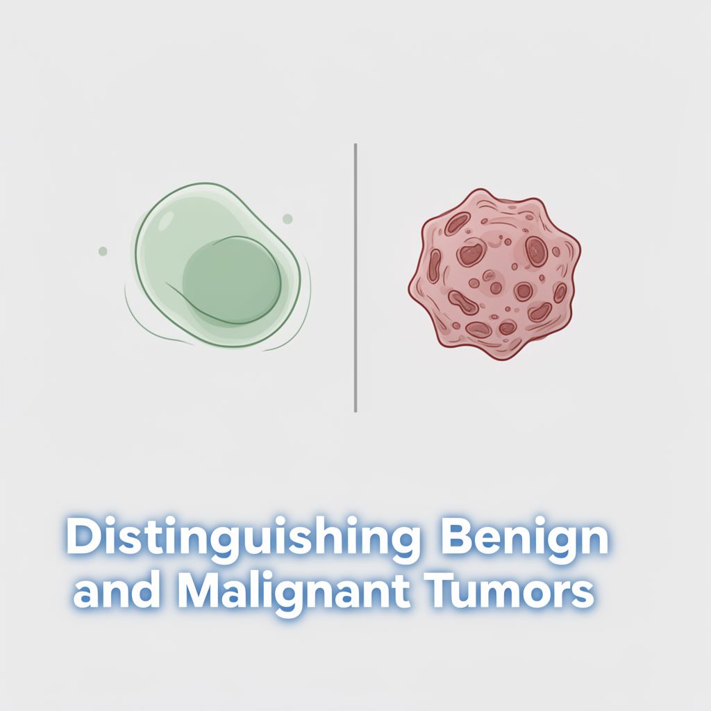
D. Benign vs. Malignant Tumors 📊
| Feature | Benign Tumor | Malignant Tumor (Cancer) | | :---------------------- | :----------------------------------------- | :------------------------------------------------- | | Spread | Localized | Local and Distant (Metastasis) | | Rate of Growth | Slow | Rapid | | Boundaries | Circumscribed, often encapsulated | Irregular, non-encapsulated | | Relationship to Tissue | Compresses surrounding normal tissues | Invades and destroys surrounding normal tissues | | Treatment | Removal will alleviate | Removal may not restore function; often requires systemic treatment |