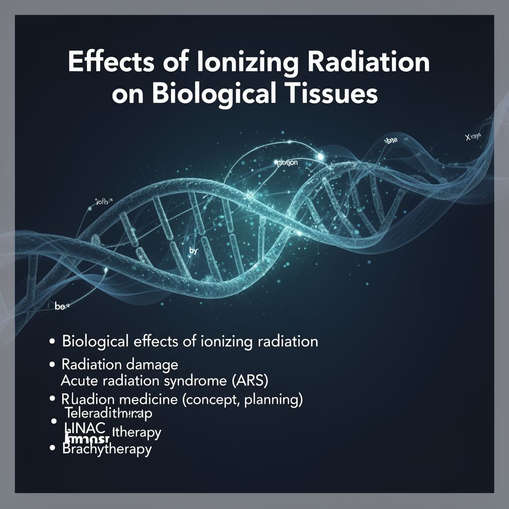
Comprehensive Study Guide: Effects of Ionizing Radiation on Biological Tissues and Therapeutic Applications
Source Information: This study material has been compiled and organized from copy-pasted text and a lecture audio transcript provided by the user.
📚 Introduction to Ionizing Radiation
Ionizing radiation plays a critical role in both understanding biological processes and in medical applications, particularly in oncology. It originates from unstable radionuclides (atoms with excess nuclear energy) and consists of subatomic particles or electromagnetic waves with sufficient energy to ionize atoms or molecules. This guide explores its fundamental aspects, impact on living organisms, and crucial role in modern medicine.
💡 Recap: Types of Ionizing Radiation
Ionizing radiation can be categorized into two main types:
- Corpuscular (Particulate) Radiation:
- Alpha (α) particles
- Beta (β) particles
- Protons
- Neutrons
- Electromagnetic Radiation:
- X-rays
- Gamma (γ) rays
Ionization can be primary (caused directly by ionizing particles) or secondary (caused by electrons released during primary ionization).
🔬 Biological Effects of Ionizing Radiation
Ionizing radiation profoundly impacts biological systems by causing chemical and biological changes at cellular and subcellular levels. This can lead to cell mutation or cell death (apoptosis or necrosis).
1️⃣ Mechanisms of Damage
The process of radiation damage unfolds over various timescales:
- Physical (10⁻¹³ s): Ionization occurs.
- Physico-chemical (10⁻¹⁰ s): Intermolecular energy transfers.
- Chemical (10⁻⁶ s): Damage to biological structures.
- Biological (seconds to years): Long-term damage at the biological level.
Damage can occur through two primary mechanisms:
- Direct Effects:
- Ionization energy is absorbed directly by critical cellular structures, such as the cytoskeleton or DNA.
- Altered chemical bonds lead to inactivation or decay of molecules.
- Most prominent in cells with low water content.
- Indirect Effects:
- More common, especially in biological tissues rich in water.
- Lysis (splitting) of water molecules leads to the production of highly reactive free radicals (e.g., H⁺, OH⁻).
- These free radicals then react with vital molecules like DNA, causing damage or mutations.
- The extent of indirect effects depends heavily on the water content of the tissue.
2️⃣ Factors Influencing Tissue Sensitivity
The biological effects of radiation depend on the dose and characteristics of the tissue. The equivalent dose is crucial and depends on the radiation type.
- Cell Differentiation Rate:
- Highly sensitive cells: Those with a high differentiation rate (rapidly dividing cells) are more susceptible. Examples include bone marrow, intestinal lining, lymphatic system, and male gonads.
- Less sensitive cells: Those with a low differentiation rate (slowly dividing or non-dividing cells) are less affected. Examples include muscles and brain tissue.
- Oxygen Levels (Oxygen Inherency): Tissues with higher levels of oxygen are generally more radiosensitive. This is because oxygen can enhance the formation of damaging free radicals.
3️⃣ Types of Radiation Effects
Radiation effects are broadly classified into two categories:
- Deterministic Effects:
- These effects are predictable and always lead to the same outcome once a certain threshold dose is exceeded.
- The severity of the effect increases with the dose.
- Caused by large-scale cell death, leading to conditions like radiation sickness.
- Examples: Hair loss, skin burns, organ failure.
- Stochastic Effects:
- These effects are unpredictable and occur randomly, with the probability of occurrence increasing with dose, but without a clear threshold.
- They result from DNA mutations that are not successfully repaired.
- Concerns about DNA:
- Potentially permanent effects (mutations).
- Can lead to cancer (somatic effects).
- Can be heritable (germline effects), affecting future generations.
- Linear No-Threshold Model: Suggests that any dose of radiation, no matter how small, carries some risk, and there is no "safe" dose. Higher doses lead to a higher risk.
- Adaptive Response: Low doses of radiation can sometimes trigger repair processes, leading to increased cellular resilience over time.
4️⃣ Lethal Dose (LD50)
The Lethal Dose 50 (LD50) indicates the dose at which 50% of exposed subjects will die within 30 days. It varies significantly across species:
- Human: 4-5 Gy
- Dog: 3 Gy
- Mouse/Rat: 7-10 Gy
- Insect: 50-100 Gy
- Note: 1 rem = 10 mSv
⚠️ Radiation Syndromes
1️⃣ Acute Radiation Syndrome (ARS)
Also known as radiation poisoning or sickness, ARS refers to effects appearing within 24 hours of exposure to high levels of radiation (typically >1 Gy).
- Symptoms depend on:
- Dose (Gy) and dose rate (Gy/s)
- Geometry of the source and location in the body
- Age, sex, and overall health status
- Manifestations:
- Lower Doses: Gastrointestinal effects (vomiting, nausea), lowered blood count (leading to infection, bleeding).
- High Doses: Neurological effects, disorientation, confusion, impaired coordination, convulsions, and potentially death within hours to days.
- Specific Forms:
- Hematologic Form: Occurs after whole-body irradiation of 1-6 Gy. Recovery in 6-8 weeks for smaller doses.
- Gastrointestinal Form: Around 10 Gy to the GIT. Symptoms (bloody diarrhea, impaired water/mineral management) appear in 4-6 days.
- Neuropsychological Form: Tens of Gy. Leads to rapid neurological decline and death.
- Skin Damage: From 3 Gy upwards, can cause hair loss (regrowth in 3 weeks if limited).
- Treatment: Blood transfusions, antibiotics.
2️⃣ Chronic Radiation Syndrome
These are long-term effects that develop over years after exposure.
- Chronic Radiation Dermatitis:
- Threshold dose: 30 Gy.
- Can manifest as atrophic (thin skin, refractive nails) or hypertrophic (keratinization, ulcers) forms. Historically seen in radiologists.
- Lens Opacity (Cataract):
- Threshold dose: 2 Gy.
- Has a long latency period, appearing years after exposure.
🏥 Radiotherapy: Principles and Applications
Radiotherapy is a medical specialization that uses the biological effects of ionizing radiation for therapeutic purposes, primarily in radiation oncology.
📊 Cancer Staging (TNM System)
Cancer staging is the process of determining the extent of cancer spread. The TNM system (Tumor, Node, Metastasis) is commonly used (e.g., T3N1M0, T2N0).
📈 Radiotherapy Diagnostics
Diagnostics are crucial for effective radiotherapy:
- Primary Cancer Diagnostic: Detection, localization, and volume assessment of primary tumors and metastases (e.g., RTG, sonography, scintigraphy (SPECT, PET), MRI).
- Cancer Therapy Planning Diagnostic: Accurate tumor localization and volume determination, prediction of therapy response (e.g., CT, NMRI combined with PET).
- Biological Response to Therapy Monitoring: Assessing therapy efficiency and predicting outcomes (e.g., CT or MRI for tumor volume changes).
✅ Mechanism and Goal of Radiotherapy
- Mechanism of Action: Based on damaging the DNA of cancer cells, either directly or indirectly through free radicals (hydroxyl radicals: H⁺, OH⁻) produced from water ionization.
- Goal: To eliminate cancer stem cells, stop tumor growth, and prevent regeneration, while minimizing damage to healthy surrounding tissues.
🎯 Therapeutic Types
Radiotherapy methods can be classified by their goal and by their technique:
- By Goal:
- Curative: Aims to completely cure the cancer.
- Palliative: Aims to slow cancer progression and alleviate symptoms.
- Adjuvant: Given after primary treatment (e.g., surgery) to reduce recurrence risk.
- Neoadjuvant: Given before primary treatment (e.g., surgery) to shrink the tumor or reduce its activity.
- By Method:
- Teleradiotherapy (External Beam Radiotherapy - EBRT): Radiation delivered from an external source.
- Brachytherapy: Short-range exposure directly to the target tissue using internal sources.
- Radioisotope Therapy: Application of radionuclides in chemical form, which are absorbed by tumor cells.
💡 Radiotherapy Planning
Computer planning is essential to ensure maximum damage to the tumor while minimizing harm to non-tumor tissues. This involves planning the appropriate external beam or internal brachytherapy treatment technique.
🚀 Teleradiotherapy (External Beam Radiotherapy)
Teleradiotherapy involves delivering radiation from a source outside the body.
📊 Dose vs. Depth Curves & Bragg Peak
- Dose Distribution: The penetration depth of radiation depends on the beam's properties (particle type, energy).
- Proton Beam (Bragg Effect/Bragg Peak): Protons release most of their energy at a specific, well-defined depth, creating a sharp peak of dose deposition known as the Bragg Peak. This allows for highly targeted dose delivery, sparing tissues beyond the tumor.
🛠️ Teleradiotherapy Techniques
- Conform Radiotherapy: The irradiated volume is precisely adapted to the irregular shape of the target tumor.
- Intensity Modulated Radiation Therapy (IMRT): The radiation beam is not only shaped but also modulated in intensity using multi-leaf collimators (MLC), allowing for highly customized dose distributions.
- Stereotactic Irradiation: Characterized by a huge dose gradient, delivering a high dose only to the target zone with a rapid fall-off in surrounding areas.
- Used for intracranial or extracranial tumors.
- Requires accurate localization using 3D coordinate systems and imaging (CT/MRI).
- Patient fixation is crucial, using invasive stereotactic frames or non-invasive masks.
🤖 Teleradiotherapy Devices
- Leskell's Gamma Knife:
- An integrated system for stereotactic radiosurgery, primarily for brain diseases (malignant/benign tumors, vascular malformations).
- Uses focused gamma radiation from multiple (e.g., 201) radioactive sources (typically ⁶⁰Co, ~1.25 MeV).
- Each individual emitter delivers a small dose, so surrounding tissue is minimally damaged.
- The therapeutic effect occurs only at the precise point where all beams converge.
- Cyber-Knife:
- Invented in the 1990s, uses an X-ray source mounted on a modified industrial robot, offering high mobility.
- Combines with real-time imaging for online correction of emitters, even with patient movement (e.g., breathing).
- Accuracy is about 1 mm.
- Often used for lung tumors and metastases.
- Does not require a stereotactic frame or anesthesia, and procedures can be outpatient.
- Linear Accelerator (LINAC):
- Electromagnetically accelerates electrons to generate bremsstrahlung (X-rays) in a target.
- Produces gamma radiation up to 500 keV (electrons up to 18 MeV).
- Used in Isocentric Radiotherapy, where beams from several directions converge at a focal center (isocenter) within the tumor, efficiently irradiating the tumor center while minimizing dose to surrounding tissues.
- Proton Therapy:
- Uses protons (hydrogen nuclei) for irradiation.
- Leverages the Bragg Peak effect to release most energy precisely at the target, with minimal exit dose.
- The beam is shaped electromagnetically, and the depth of the Bragg Peak is controlled by beam energy.
🩹 Brachyradiotherapy (Internal Radiotherapy)
Brachyradiotherapy is a local radiotherapy method where the radiation source is placed in close contact with the tumor.
1️⃣ Closed Radioisotopes
- Used for small tumor volumes.
- The radiation source is sealed and inserted by implantation or tapping directly into or near the tumor.
- Requires mechanical accessibility of the target lesion.
- Can be temporary or permanent.
- Common sources: ²²⁶Ra, ⁶⁰Co, ¹⁹²Ir.
2️⃣ Open Radioisotopes
- Based on selective radionuclide compounds that are captured and accumulated inside tumor cells.
- The radiation eliminates the tumor from within.
- Mostly uses beta (β) decaying radionuclides due to their small range of radiation (e.g., ~4 mm), minimizing damage to distant healthy tissues.
- Monitored with scintigraphy.
- Optimal dosing balances maximal radiation effect with minimal radiotoxicity to target tissue.
🧪 Examples of Open Radioisotopes:
- Thyroid Glands (¹³¹I):
- Thyroid tumor tissue selectively absorbs and accumulates iodine.
- Applied orally, with minimal effect on other body tissues.
- ¹³¹I: Activity 3-7 GBq, half-life 8 days, emits β particles (606 keV) and γ rays (284 keV).
- Hematology (³²P):
- Used for treating polycythemia vera (abnormal production of red blood cells).
- Radioactive phosphorus is absorbed by bone marrow, where β particles impact blood stem cells.
- Major disadvantage: risk of acute leukemia.
- ³²P: Activity 200-500 MBq, half-life 14.3 days, emits β particles (1.7 MeV).
- Bone (⁸⁹Sr):
- Has similar metabolism to calcium, accumulating significantly more in metastatic bone than in healthy bone.
- Used for pain relief in bone metastases.
- ⁸⁹Sr: Activity 150 MBq, half-life 50.5 days, emits β particles (1.5 MeV).