Upper Limb Practical Exam Study Guide
Source Information: This study material is compiled from a lecture audio transcript on preparing for an upper limb practical exam.
Introduction: Navigating Your Upper Limb Practical Exam 💡
This guide is designed to help you prepare effectively for your upper limb practical exam. It breaks down the essential anatomical structures and concepts that are frequently tested, providing a clear roadmap for your study. By focusing on these key areas, you'll gain a comprehensive understanding and boost your confidence for the exam.
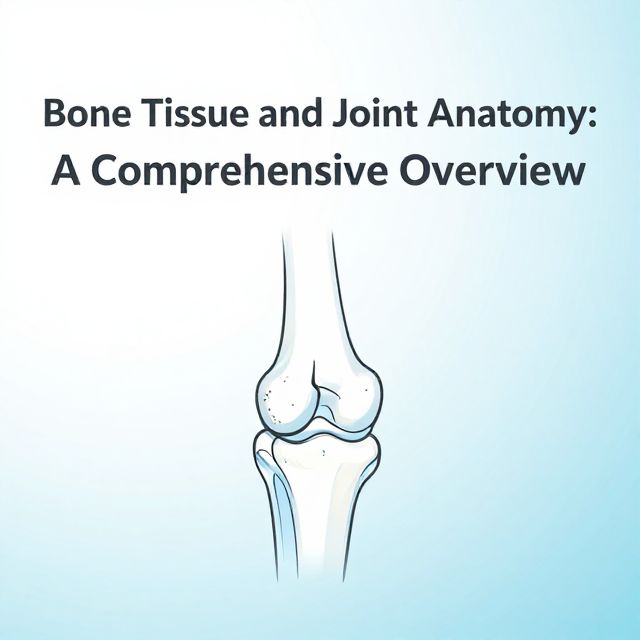
1. Bones and Joints: The Foundation of Movement 🦴
Understanding the skeletal framework and how it articulates is fundamental.
1.1. Bones: Identification and Key Landmarks ✅
You must be able to identify all major bones of the upper limb and their significant landmarks.
- Shoulder Girdle: Scapula, Clavicle
- Arm: Humerus
- Forearm: Radius, Ulna
- Hand: Carpals, Metacarpals, Phalanges
Key Landmarks to Know:
- Scapula: Acromion process, spine, coracoid process, glenoid cavity.
- Humerus: Greater tubercle, lesser tubercle, deltoid tuberosity, medial and lateral epicondyles, capitulum, trochlea.
- Radius: Radial head, neck, tuberosity, styloid process.
- Ulna: Olecranon process, coronoid process, trochlear notch, radial notch, styloid process.
- Carpals: Identify individual carpal bones (e.g., scaphoid, lunate, triquetrum, pisiform, trapezium, trapezoid, capitate, hamate).
1.2. Joints: Type, Movement, and Stability 📚
For each major joint, understand its classification, the movements it permits, and the primary ligaments that stabilize it.
- Shoulder Joint (Glenohumeral):
- Type: Ball and socket.
- Movements: Flexion, extension, abduction, adduction, internal rotation, external rotation, circumduction.
- Stability: Primarily provided by the rotator cuff muscles (SITS: Supraspinatus, Infraspinatus, Teres minor, Subscapularis) and glenohumeral ligaments.
- Elbow Joint:
- Type: Hinge (humeroulnar, humeroradial) and pivot (proximal radioulnar).
- Movements: Flexion, extension (humeroulnar/humeroradial); pronation, supination (radioulnar).
- Stability: Ulnar collateral ligament, radial collateral ligament, annular ligament.
- Wrist Joint (Radiocarpal):
- Type: Condyloid.
- Movements: Flexion, extension, radial deviation, ulnar deviation, circumduction.
- Stability: Palmar and dorsal radiocarpal ligaments, ulnar collateral ligament, radial collateral ligament.
- Joints of the Hand:
- Intercarpal, Carpometacarpal, Metacarpophalangeal, Interphalangeal joints: Know their types and primary movements (e.g., flexion/extension, abduction/adduction).
Practical Focus: 1️⃣ Be prepared to identify bones and landmarks on models or images. 2️⃣ Demonstrate and explain the movements allowed at each joint.
2. Muscles and Nerves: The Movers and Shakers 💪
These systems are responsible for all upper limb function and sensation.
2.1. Muscles: Origin, Insertion, Action, and Innervation ✅
Group muscles by region and understand their key characteristics.
- Shoulder Girdle Muscles:
- Examples: Trapezius, Serratus Anterior, Levator Scapulae, Rhomboids.
- Focus: Their role in scapular movement and stability.
- Muscles Acting on the Shoulder Joint:
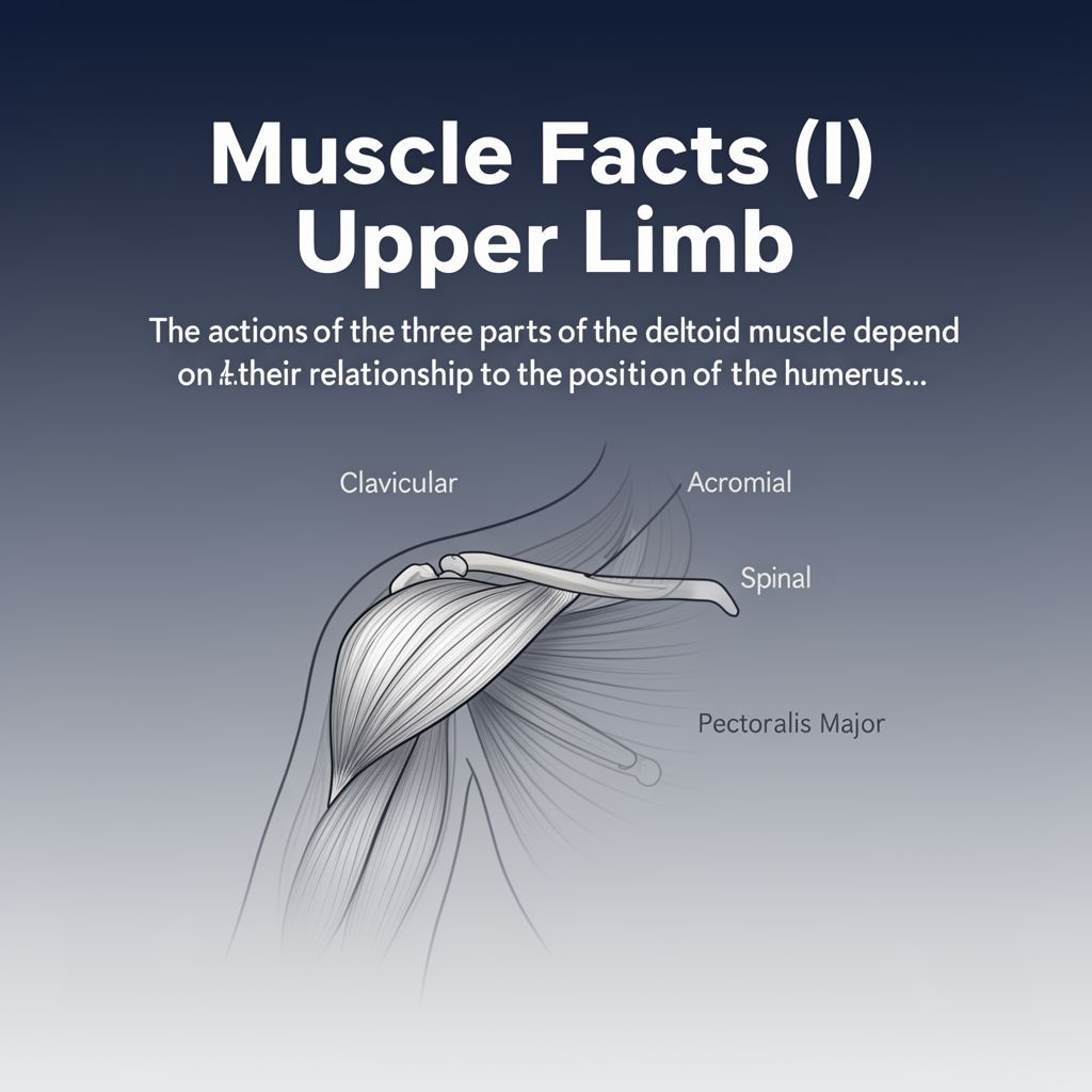
- Examples: Deltoid, Pectoralis Major, Latissimus Dorsi.
- Rotator Cuff (SITS): Supraspinatus (abduction), Infraspinatus (external rotation), Teres minor (external rotation), Subscapularis (internal rotation).
- Muscles of the Arm:
- Anterior Compartment (Flexors): Biceps Brachii, Brachialis, Coracobrachialis.
- Posterior Compartment (Extensors): Triceps Brachii.
- Muscles of the Forearm:
- Anterior Compartment (Flexors/Pronators): Superficial and deep layers (e.g., Flexor Carpi Radialis, Flexor Carpi Ulnaris, Palmaris Longus, Flexor Digitorum Superficialis/Profundus, Pronator Teres).
- Posterior Compartment (Extensors/Supinators): Superficial and deep layers (e.g., Extensor Carpi Radialis Longus/Brevis, Extensor Carpi Ulnaris, Extensor Digitorum, Supinator).
- Intrinsic Muscles of the Hand:
- Thenar Eminence: Muscles of the thumb (e.g., Abductor Pollicis Brevis, Flexor Pollicis Brevis, Opponens Pollicis).
- Hypothenar Eminence: Muscles of the little finger (e.g., Abductor Digiti Minimi, Flexor Digiti Minimi Brevis, Opponens Digiti Minimi).
- Other: Lumbricals, Interossei (Palmar and Dorsal).
Practical Focus: 1️⃣ Be able to palpate muscles and demonstrate their actions. 2️⃣ Know the innervation for major muscle groups.
2.2. Nerves: Course, Innervation, and Sensory Distribution ⚠️
Focus on the brachial plexus and its major terminal branches.
- Brachial Plexus: Understand its general organization (Roots, Trunks, Divisions, Cords, Branches).
- Five Major Terminal Nerves:
- Musculocutaneous Nerve:
- Course: Pierces coracobrachialis, runs between biceps and brachialis.
- Motor: Innervates anterior arm muscles (biceps, brachialis, coracobrachialis).
- Sensory: Lateral forearm.
- Axillary Nerve:
- Course: Passes through quadrangular space.
- Motor: Deltoid, Teres Minor.
- Sensory: "Regimental badge" area over deltoid.
- Radial Nerve:
- Course: Runs in radial groove of humerus, then anterior to lateral epicondyle.
- Motor: All posterior arm and forearm muscles (extensors, supinators).
- Sensory: Posterior arm, forearm, and dorsum of hand (lateral 3.5 digits).
- Clinical Relevance: "Wrist drop" if damaged.
- Median Nerve:
- Course: Runs with brachial artery, passes through carpal tunnel.
- Motor: Most anterior forearm muscles (flexors, pronators), thenar muscles, first two lumbricals.
- Sensory: Lateral palm, palmar aspect of lateral 3.5 digits.
- Clinical Relevance: Carpal Tunnel Syndrome.
- Ulnar Nerve:
- Course: Passes posterior to medial epicondyle ("funny bone"), then through Guyon's canal at wrist.
- Motor: Flexor Carpi Ulnaris, medial half of Flexor Digitorum Profundus, most intrinsic hand muscles (hypothenar, interossei, 3rd & 4th lumbricals).
- Sensory: Medial palm, palmar and dorsal aspect of medial 1.5 digits.
- Clinical Relevance: "Claw hand" if damaged.
- Musculocutaneous Nerve:
3. Blood Vessels and Surface Anatomy: Supply Lines and Landmarks 📊
These areas cover the vascular supply and how to identify structures externally.
3.1. Blood Vessels: Major Arteries and Veins 📈
Trace the major vessels and understand their distribution.
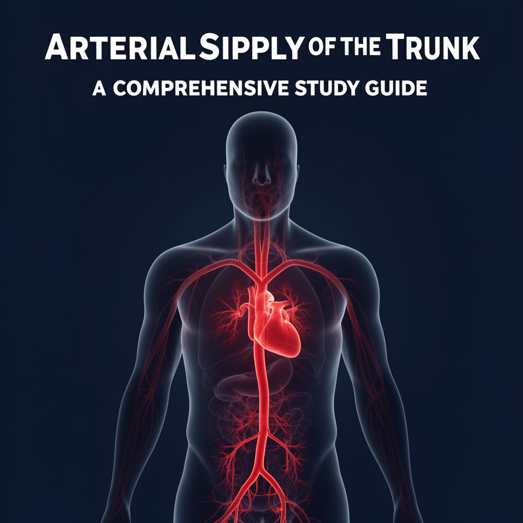
- Arteries:
- Subclavian Artery → Axillary Artery → Brachial Artery → Radial Artery & Ulnar Artery → Palmar Arches (Superficial and Deep).
- Veins:
- Superficial: Cephalic Vein, Basilic Vein, Median Cubital Vein.
- Deep: Accompanying arteries (e.g., brachial veins, radial veins, ulnar veins).
3.2. Surface Anatomy: Palpation and Visual Identification 🗺️
Be able to locate and identify structures by touch and sight.
- Key Palpable Landmarks:
- Clavicle, Acromion, Spine of Scapula.
- Greater Tubercle of Humerus.
- Medial and Lateral Epicondyles of Humerus.
- Olecranon Process.
- Radial and Ulnar Styloid Processes.
- Head of Radius.
- Metacarpals and Phalanges.
- Key Regions/Structures:
- Cubital Fossa: Boundaries and contents (Median nerve, Brachial artery, Biceps tendon).
- Anatomical Snuffbox: Boundaries and contents (Radial artery, Scaphoid bone).
- Pulse Points: Radial pulse, Brachial pulse.
Practical Focus: 1️⃣ Practice palpating these landmarks on yourself or a study partner. 2️⃣ Be able to identify regions like the cubital fossa and anatomical snuffbox.
Your Path to Practical Exam Success 🚀
To excel in your upper limb practical exam, remember these key strategies:
- Master the Basics: Solidify your knowledge of bones, joints, muscles, and nerves.
- Understand Function: Connect structure to function – what movements do muscles produce? What sensation does a nerve provide?
- Clinical Relevance: Think about how nerve damage or muscle injury would present clinically.
- Practice Palpation: The more you feel and identify structures on a real person, the better.
- Visualize and Draw: Use anatomical models, draw diagrams, and label structures.
- Explain Out Loud: Articulate concepts to a study partner or even to yourself. This reinforces learning and identifies gaps in your understanding.
You have the tools to prepare effectively. Study smart, practice diligently, and you will succeed!