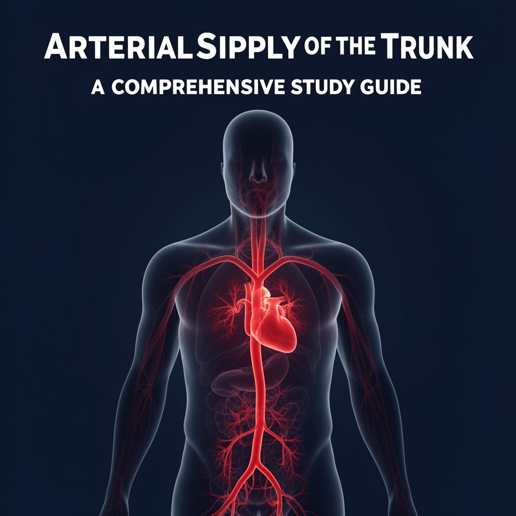
Arterial Supply of the Trunk: A Comprehensive Study Guide
Source Information: This study material has been compiled from a copy-pasted text and a lecture audio transcript.
📚 Introduction to the Aorta and its Divisions
The arterial blood supply of the human trunk is fundamentally driven by the aorta, the largest artery in the body. Originating from the left ventricle of the heart, the aorta is the primary conduit responsible for distributing oxygenated blood to the entire body. Its extensive course is divided into three main sections based on its anatomical position:
- Ascending Aorta (Aorta Ascendens): Extends from the left ventricle to the right second intercostal space.
- Aortic Arch (Arcus Aortae): Spans from the right second intercostal space to the left second intercostal space, typically at the level of the T4 vertebra.
- Descending Aorta (Aorta Descendens): Continues from the aortic arch downwards.
The descending aorta is further subdivided into two major parts:
- Thoracic Aorta (Pars Thoracica Aortae): Located within the chest cavity.
- Abdominal Aorta (Pars Abdominalis Aortae): Located within the abdominal cavity.
🫁 Thoracic Aorta (Pars Thoracica Aortae)
The thoracic aorta is the segment of the descending aorta that extends from the T4 to the T12 vertebral levels. Initially positioned to the left of the vertebral column, it gradually approaches the midline as it descends, becoming centrally located at the aortic hiatus.
This section of the aorta is responsible for supplying blood to the thoracic organs (excluding the heart itself) and the chest wall. Its branches are categorized into visceral and parietal.
✅ Visceral Branches of the Thoracic Aorta
These branches supply the organs within the thoracic cavity:
- Bronchial Arteries (Rr. Bronchiales): 4-5 thin branches that are the nutritive arteries for the lungs.
- Esophageal Arteries (Rr. Oesophageales): 4-5 thin branches arising from the anterior surface of the aorta, supplying the esophagus.
- Pericardial Arteries (Rr. Pericardiaci): Small branches distributed on the posterior surface of the pericardium.
- Mediastinal Arteries (Rr. Mediastinales): Small, thin branches supplying lymph nodes and connective tissue within the mediastinum.
✅ Parietal Branches of the Thoracic Aorta
These branches supply the walls of the thoracic cavity:
- Posterior Intercostal Arteries (Aa. Intercostales Posteriores):
- Nine pairs (3rd-11th) arise directly from the thoracic aorta.
- The 1st and 2nd posterior intercostal arteries originate from the superior intercostal artery (a. intercostalis suprema), which is a branch of the costocervical trunk (truncus costocervicalis) from the subclavian artery.
- These arteries travel within their respective intercostal spaces, typically in the costal groove.
- They anastomose with the anterior intercostal arteries (branches of the internal thoracic artery or musculophrenic artery).
- 💡 Clinical Note: Within the intercostal spaces, the arrangement of structures from superior to inferior is VAN (Vein, Artery, Nerve).
- Branches of Posterior Intercostal Arteries:
- Dorsal Ramus (R. Dorsalis): Passes between the necks of adjacent ribs, gives off a spinal ramus (r. spinalis) that enters the vertebral canal to supply the spinal cord, its meninges, and vertebrae. It also supplies back muscles and skin.
- Collateral Ramus (R. Collateralis): Arises near the costal angle, descends to the superior border of the rib below, and anastomoses with the anterior intercostal artery.
- Lateral Cutaneous Ramus (R. Cutaneus Lateralis): Supplies the skin.
- Lateral Mammary Rami (Rr. Mammarii Laterales): Arise from the 3rd-5th intercostal arteries, supplying the lateral part of the breast.
- Muscular Rami (R. Muscularis): Supply intercostal, pectoral, and serratus anterior muscles.
- Subcostal Artery (A. Subcostalis): Arises from the thoracic aorta and runs inferior to the 12th rib.
- Superior Phrenic Artery (A. Phrenica Superior): Arises from the inferior end of the thoracic aorta and distributes to the diaphragm.
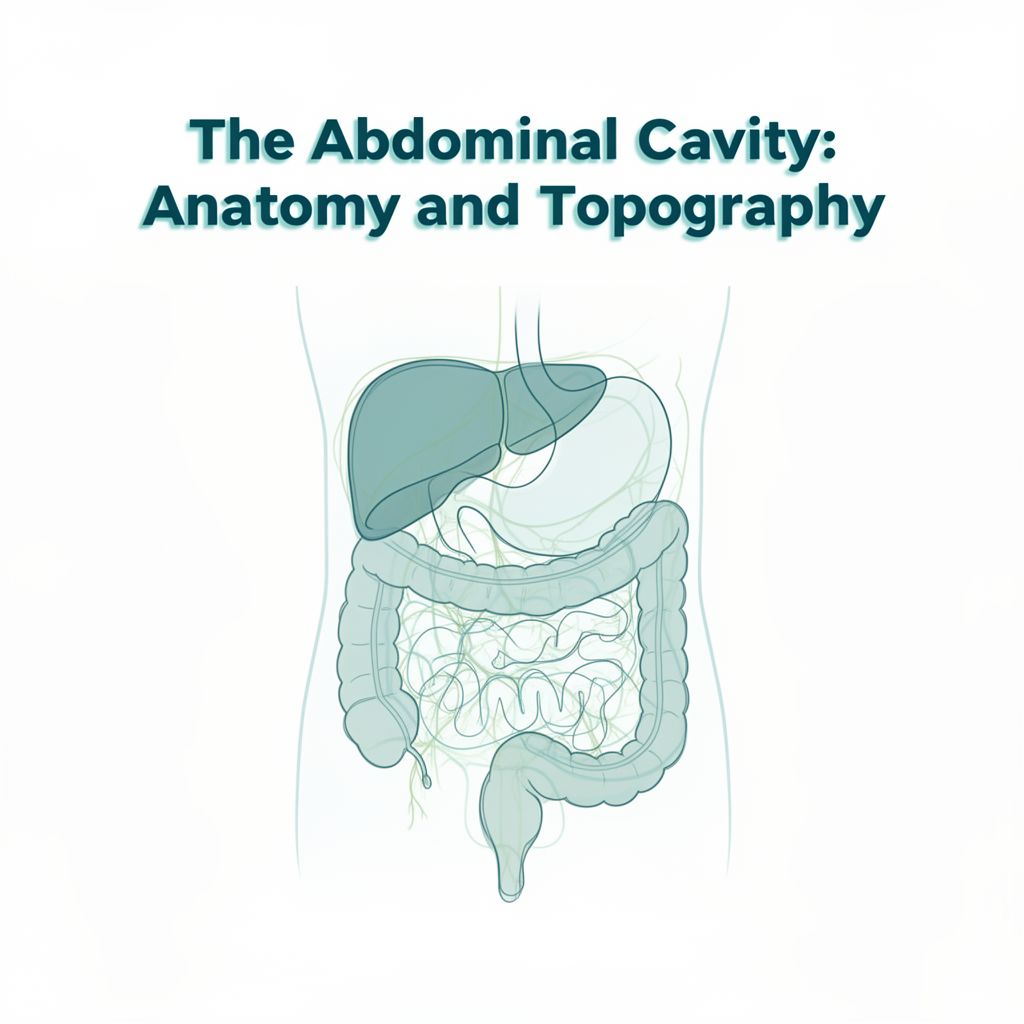
🍎 Abdominal Aorta (Pars Abdominalis Aortae)
The abdominal aorta begins after the thoracic aorta passes through the aortic hiatus of the diaphragm (at T12). It extends from T12 to L4, where it terminates by bifurcating into the right and left common iliac arteries. It lies anterior to the vertebral column in the abdominal cavity.
The abdominal aorta supplies both the abdominal wall (parietal branches) and the abdominal organs (visceral branches).
📊 Visceral Branches of the Abdominal Aorta
These branches supply the organs within the abdominal cavity. They are divided into single (unpaired) branches and paired branches.
1️⃣ Single (Unpaired) Visceral Branches
These typically arise from the anterior surface of the aorta and primarily supply organs of the digestive system and the spleen.
- Celiac Trunk (Truncus Coeliacus):
- Arises just below the aortic hiatus.
- A short trunk that quickly divides into three main branches:
- Left Gastric Artery (A. Gastrica Sinistra): The smallest branch, supplies the lower esophagus (via rr. oesophageales) and the lesser curvature of the stomach. It anastomoses with esophageal branches from the thoracic aorta.
- Common Hepatic Artery (A. Hepatica Communis): Supplies the liver, stomach, duodenum, and pancreas. It gives rise to:
- Proper Hepatic Artery (A. Hepatica Propria): Enters the liver, gives off the right gastric artery (a. gastrica dextra) and cystic artery (a. cystica) for the gallbladder.
- Gastroduodenal Artery (A. Gastroduodenalis): Passes behind the first part of the duodenum, giving off the superior pancreaticoduodenal artery (a. pancreaticoduodenalis superior) and the right gastro-omental (epiploic) artery (a. gastro-omentalis dextra).
- Splenic Artery (A. Lienalis): The largest branch of the celiac trunk, tortuous in its course along the superior border of the pancreas. Supplies the pancreas (via rr. pancreatici), spleen, and stomach (via short gastric arteries aa. gastrici breves and left gastro-omental artery a. gastro-omentalis sinistra).
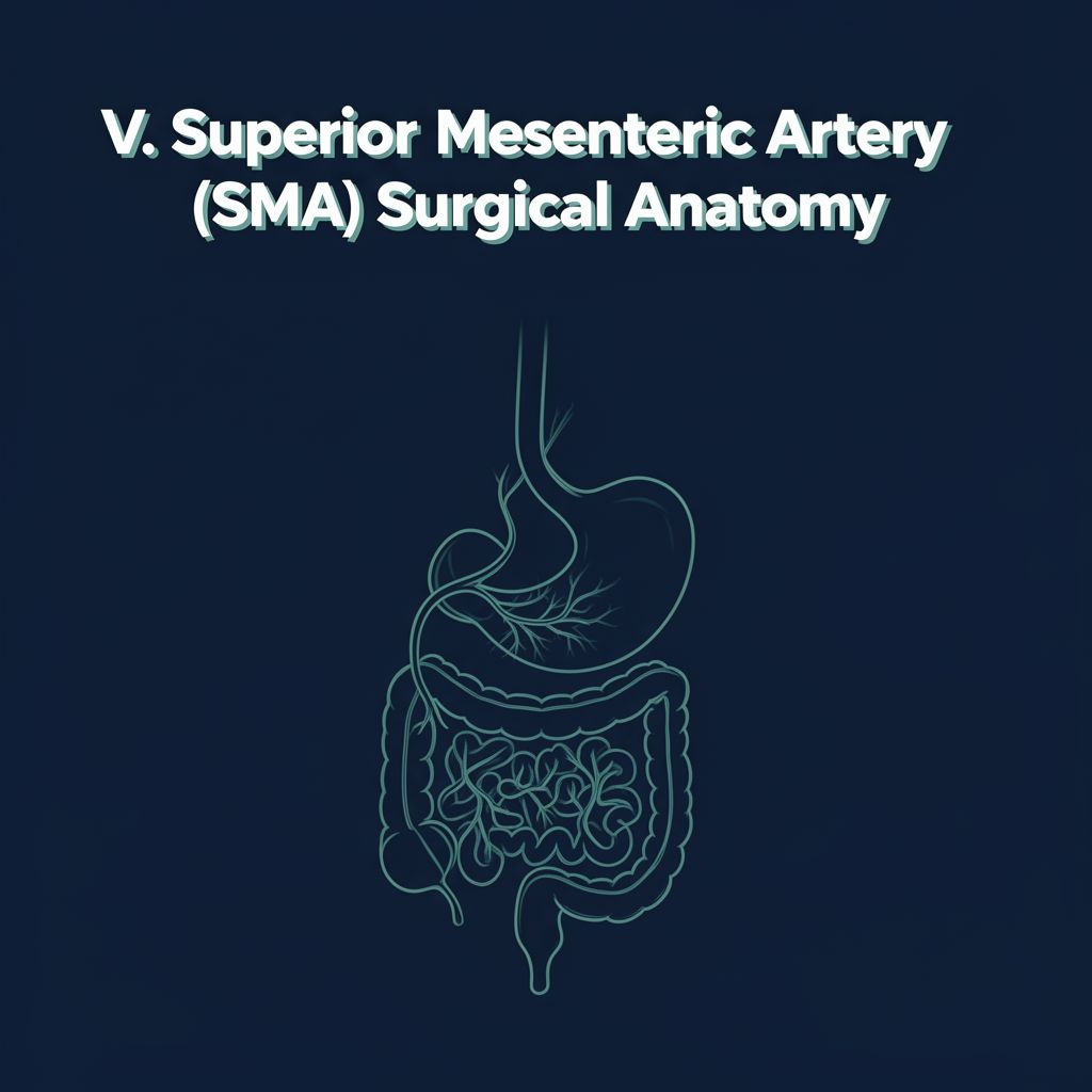
- Superior Mesenteric Artery (A. Mesenterica Superior - AMS):
- Arises below the celiac trunk.
- Supplies the lower duodenum, jejunum, ileum, cecum, ascending colon, and the right two-thirds of the transverse colon.
- Key Branches:
- Inferior Pancreaticoduodenal Artery (A. Pancreaticoduodenalis Inferior): Supplies the head of the pancreas and adjacent duodenum, anastomosing with the superior pancreaticoduodenal artery.
- Jejunal and Ileal Arteries (Aa. Jejunales et Aa. Ileales): 12-15 arteries supplying the jejunum and ileum, forming arterial arcades within the mesentery.
- Ileocolic Artery (A. Ileocolica): Supplies the terminal ileum, cecum (anterior and posterior cecal arteries), and appendix (appendicular artery).
- Right Colic Artery (A. Colica Dextra): Supplies the ascending colon, anastomosing with the ileocolic and middle colic arteries.
- Middle Colic Artery (A. Colica Media): Supplies the right two-thirds of the transverse colon, anastomosing with the right colic and left colic arteries to form the marginal artery of the colon (of Drummond).
- Inferior Mesenteric Artery (A. Mesenterica Inferior - AMI):
- Supplies the left one-third of the transverse colon, descending colon, sigmoid colon, and the superior part of the rectum.
- Key Branches:
- Left Colic Artery (A. Colica Sinistra): Supplies the left one-third of the transverse colon and descending colon, anastomosing with the middle colic artery and sigmoid arteries.
- Sigmoid Arteries (Aa. Sigmoideae): 2-3 arteries supplying the sigmoid colon, forming anastomoses with each other and with the left colic and superior rectal arteries.
- Superior Rectal Artery (A. Rectalis Superior): The terminal branch of the AMI, supplies the superior part of the rectum, anastomosing with the middle and inferior rectal arteries.
2️⃣ Paired Visceral Branches
These typically arise from the lateral surfaces of the aorta and supply non-digestive organs.
- Middle Suprarenal Arteries (A. Suprarenalis Media): A pair of small arteries supplying the adrenal (suprarenal) glands. They anastomose with superior (from inferior phrenic) and inferior (from renal) suprarenal arteries.
- Renal Arteries (A. Renalis): The thickest branches of the abdominal aorta, arising at the level of L1-L2. They supply the kidneys. The right renal artery passes behind the inferior vena cava.
- Testicular/Ovarian Arteries (A. Testicularis / A. Ovarica):
- Arise inferior to the renal arteries.
- Testicular Arteries: Descend retroperitoneally, cross the ureters and external iliac arteries, enter the inguinal canal, and supply the testes and epididymis.
- Ovarian Arteries: Descend into the pelvis, supply the ovaries, uterine tubes, and ureters. They anastomose with the uterine artery.
📈 Parietal Branches of the Abdominal Aorta
These branches supply the walls of the abdominal cavity.
- Paired Parietal Branches:
- Inferior Phrenic Arteries (A. Phrenica Inferior): A pair of arteries supplying the diaphragm, often arising just above the celiac trunk. They give off superior suprarenal arteries (aa. suprarenales superiores).
- Lumbar Arteries (Aa. Lumbales): Four pairs of arteries arising from the posterior surface of the aorta, corresponding to the first four lumbar vertebrae. They supply the abdominal wall, back muscles, joints, and skin (via r. dorsalis), and the spinal cord, meninges, and vertebrae (via r. spinalis). They form anastomoses with intercostal, subcostal, iliolumbar, deep circumflex iliac, and inferior epigastric arteries.
- Single Parietal Branch:
- Median Sacral Artery (A. Sacralis Mediana): Arises from the posterior aspect of the aorta just above its bifurcation. It descends along the midline over the L4-L5 vertebrae, sacrum, and coccyx, giving off small branches to the rectum and anastomosing with iliolumbar and lateral sacral arteries.
🔚 Aorta Termination: Common Iliac Arteries
The abdominal aorta terminates at the level of the fourth lumbar vertebra (L4) by bifurcating into its two terminal branches:
- Right Common Iliac Artery (A. Iliaca Communis Dextra)
- Left Common Iliac Artery (A. Iliaca Communis Sinistra)
These common iliac arteries descend laterally and inferiorly to the sacroiliac joint, where each divides into:
- Internal Iliac Artery (A. Iliaca Interna): Primarily supplies the pelvic organs, gluteal region, and perineum.
- External Iliac Artery (A. Iliaca Externa): Continues into the thigh as the femoral artery, supplying the lower limb.
This comprehensive arterial network ensures that all structures within the trunk and beyond receive the vital oxygenated blood necessary for their function.