This study material has been compiled from a combination of copy-pasted text and a lecture audio transcript.
Comprehensive Guide to Pulp Conditions: Inflammation, Necrosis, Degeneration, and Calcifications 🦷
This guide provides a detailed overview of various pulp conditions, including inflammatory responses like Pulpitis Polypoza, the irreversible processes of Pulp Necrosis and Gangrene, and degenerative changes such as Atrophy, Fibrosis, and Pulp Calcifications. Understanding these conditions is crucial for accurate diagnosis and effective treatment in endodontics.
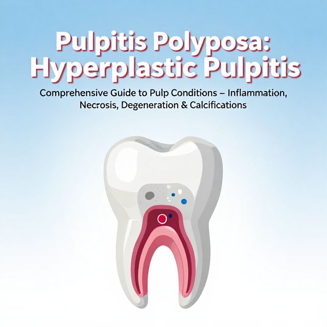
1. Pulpitis Polypoza (Chronic Hyperplastic Pulpitis) 🌿
📚 Definition: Pulpitis Polypoza, also known as Chronic Hyperplastic Pulpitis, is a chronic inflammatory condition of the pulp where granulation tissue proliferates into a carious cavity, often becoming covered by oral squamous epithelium. It typically occurs in young pulp exposed by extensive carious lesions.
✅ Key Characteristics:
- Granulation Tissue: The polyp initially consists primarily of granulation tissue, appearing as a growth extending from the pulp chamber.
- Epithelialization: The surface of the proliferating tissue is often covered by oral squamous epithelium.
- Pain Sensation: Due to a sparse distribution of nerve fibers, the polyp is usually painless upon contact.
- Bleeding: It tends to bleed easily when touched due to its rich vascular network.
- Pain on Chewing: Pain is typically experienced only during chewing, due to pressure on the tissue.
💡 Conditions for Development: For a pulp polyp to develop, specific conditions must be present:
- Open and Wide Cavity: Provides space for tissue proliferation.
- Resilient and Young Pulp: Possesses a strong regenerative capacity.
- Prolonged, Mild Irritation: Stimulates chronic inflammatory response without causing rapid necrosis.
📊 Radiographic Features:
- The periodontal ligament (PDL) space and lamina dura usually appear normal.
- In young patients, long-term mild irritation can stimulate periapical bone formation (pulpo-periapical osteosclerosis), often seen as a dense bone area around the apices of lower molars with large carious lesions.
⚠️ Differential Diagnosis: It is crucial to differentiate Pulpitis Polypoza from a gingival polyp, which originates from the gingival tissue rather than the pulp.
🩹 Treatment: Treatment involves the removal of the polyp under local anesthesia. This can be achieved through:
- Excision or Cauterization: Surgical removal or burning away of the polyp.
- Subsequent Procedures: This is followed by either pulpotomy (amputation) or root canal therapy, depending on the extent of pulp involvement and the tooth's restorability.
2. Pulp Necrosis 💀
📚 Definition: Pulp necrosis refers to the death of pulp tissue. It can result from acute or chronic inflammation, traumatic injury, or an abrupt interruption of blood circulation. It may also occur in advanced stages of pulp degeneration.
✅ Types of Necrosis: Pulp necrosis can be partial or total, depending on the extent of tissue involvement. Two main types are recognized:
-
Coagulation Necrosis:
- Mechanism: Occurs when blood supply is reduced or completely cut off.
- Appearance: The tissue transforms into a soft, cheese-like mass (caseation), composed of proteins, fats, and water.
-
Liquefaction Necrosis:
- Mechanism: Proteolytic enzymes soften and liquefy the tissues.
- Clinical Sign: Often presents with a discharge of blood and exudate from the cavity.
3. Pulp Gangrene 🦠
📚 Definition: Pulp gangrene describes a condition where infected vital pulp dies due to inflammatory processes, or when previously non-vital pulp subsequently becomes infected.
🔬 Chemistry of Gangrene: In gangrenous pulp, microorganisms completely break down the tissue. Proteins, carbohydrates, and fats undergo a series of chemical decompositions.
- Putrefaction: The decomposition of proteins by anaerobic bacteria.
- Foul Odor: This process releases various foul-smelling intermediate and final products, which are responsible for the characteristic unpleasant odor often detected when the dental cavity is accessed. These include:
- Intermediate Proteolytic Products: Indole, skatole (from tryptophan deamination), putrescine, cadaverine (from tryptophan decarboxylation), and indican.
- Final Products: Hydrogen sulfide, ammonia, water, and fatty acids.
- Bacterial Toxins: Exotoxins (secreted by bacteria) and endotoxins (released upon bacterial breakdown).
🩺 Clinical Signs:
- Pain: In cases of total pulp necrosis where periodontal tissues are unaffected, there is typically no pain or sensitivity to percussion or palpation.
- Vitality Tests: Vitality tests will yield negative results. However, in multi-rooted teeth, one root might remain vital, potentially requiring anesthesia during treatment.
- Color Change:
- Trauma-induced necrosis: May lead to a yellow-brown discoloration due to hemolysis of red blood cells.
- Gangrene: Often presents with a gray discoloration of the tooth. (Note: Some necrotic teeth may retain normal color).
📊 Radiographic Features:
- Pulp Dimensions: Teeth with long-standing gangrene may exhibit wider pulp dimensions compared to adjacent or symmetrical teeth. This is due to arrested dentin formation and decalcification of the root canal.
- Periodontal Ligament: Widening of the apical periodontal membrane is frequently observed.
🩹 Treatment: Root canal therapy for gangrenous teeth requires:
- Biomechanical Enlargement: Thorough shaping and cleaning of the root canal system.
- Calcium Hydroxide: Application of calcium hydroxide is often necessary as an intracanal medicament.
4. Pulp Degenerative Changes: Atrophy and Fibrosis 📉
Pulp tissue undergoes various degenerative changes influenced by factors such as attrition, abrasion, erosion, trauma, operative procedures, and caries. These changes include atrophic degeneration and fibrosis.
-
Atrophy:
- Definition: Refers to a reduction in the size of cells or an organ due to a decrease in the size of its constituent specialized cells.
- Pulp Manifestation: In the pulp, this means a decrease in the size of pulp cells.
-
Fibrosis:
- Definition: Characterized by an increase in mature collagen fibers per unit area.
- Pulp Manifestation: While collagen fibers increase, there is a decrease in both the number and size of pulp cells. Odontoblasts may appear flattened or cuboidal, resembling shrunken particles within a dense fibrous matrix.
📈 Accelerated Aging and its Consequences:
- Induced Aging: Excessive stimuli (e.g., severe attrition/abrasion) can accelerate the aging process, leading to:
- Dentin Formation: Increased dentin formation, narrowing the canal lumen.
- Cementum Formation: Periapical cementum formation in the apical foramen and apical canal, further narrowing the canal.
- Pulp Changes with Age (or accelerated aging):
- Progressive reduction in pulp chamber size.
- Accumulation of calcified salts, progressing from the root pulp towards the coronal pulp.
- Decrease in blood vessels and associated arteriosclerotic changes in the coronal pulp.
- Fibrotic transformation of connective tissue, affecting blood vessels and nerves.
- Loss of myelinated and unmyelinated axons, leading to decreased sensitivity.
- Physiological Implications:
- Decreased Dentin Permeability: Reduces the effect of irritants, protecting the pulp.
- Reduced Repair Potential: The pulp's ability to protect and repair itself against irritants diminishes.
5. Pulp Calcifications (Pulp Stones / Denticles) 💎
📚 Definition: Pulp calcifications are common mineralized masses found within the pulp tissue. They can occur in a single tooth, multiple teeth, primary or permanent teeth, and even in unerupted or sound teeth. They can develop at any age and in any part of the pulp tissue.
✅ Types of Pulp Calcifications:
-
Dystrophic Calcification:
- Formation: Accumulation of calcium salts within dead or degenerated tissue.
- Mechanism: Local alkalinity in damaged tissues attracts these salts. Can occur in young pulps affected by circulatory disturbances, blood clots, or around single degenerated cells.
-
Diffuse Calcification:
- Appearance: Characterized by numerous small and irregular calcium deposits scattered throughout the pulp.
-
Denticles / Pulp Stones:
- Appearance: Large, concentric clusters of calcium deposits.
- Classification by Localization:
- Free: Found freely within the pulp chamber.
- Embedded: Surrounded by dentin.
- Attached: Adhering to the dentin wall.
- Classification by Structure:
- True Denticle: Contains tubular orthodentin, resembling normal dentin. Formed during odontogenesis through epithelio-mesenchymal interactions, typically near the furcation or root sheath.
- False Denticle: Composed of concentric layers of calcified material that does not resemble dentin.
- Diffuse Calcifications: Small, irregular calcified deposits within the pulp tissue.
- Formation: Pulp stones can form at any time and location within the pulp from the calcification of isolated pulp structures. Their size can range from microscopic particles to masses large enough to fill the entire pulp chamber.
- Composition: Can consist of normal dentin (ortho-dentin), non-tubular (fibrous) dentin, or irregular calcified material.
- Etiology: Delayed eruption can predispose to pulp calcification.
⚙️ Mechanisms of Pulp Calcification:
- Calcification of Tissue Components:
- An initial calcification (e.g., collagen fibril, ground substance, or necrotic cell debris) acts as a nucleus for the concentric lamellar or radial deposition of other calcified material.
- This process can be triggered by an imbalance between promoters and inhibitors of calcification, or local damage to inhibitors.
- Accumulation of calcium salts in areas of hyaline degeneration, where electrolyte balance is disturbed, preventing calcium from remaining in solution.
- Epithelio-Mesenchymal Interactions: Primarily involved in the formation of true denticles during tooth development.
🔬 Tissues Forming Calcifications:
- Orthodentin (Tubular Dentin): Found in both denticles and pulp stones. In true denticles, odontoblasts line the periphery, but their height decreases, and they eventually die as the denticle grows.
- Regular Calcified Material: Can be found in the peripheral layers of both pulp stones and denticles.
- Irregular Calcified Material: Often found at the center of pulp stones, sometimes in lamellar formations on their surface, and even on the surface of denticles. These can grow to considerable sizes by mineral addition to adjacent matrix fibrils.
6. Clinical Significance and Treatment of Calcified Canals ⚠️
💡 Clinical Importance of Pulp Calcifications:
- Dental Neuralgia: Some studies suggest pulp calcifications can contribute to dental neuralgia.
- Obstruction: Calcifications in the pulp chamber can obstruct the entry to the root canal, making canal localization difficult.
- Root Canal Treatment (RCT) Challenges: Calcifications in the root pulp can prevent complete instrumentation and obturation up to the physiological foramen, increasing the risk of treatment failure.
🩹 Considerations for RCT in Calcified Canals: Performing root canal treatment on teeth with calcified canals requires meticulous technique:
- Slow and Gentle Instrumentation: Canal instruments should be used slowly and without excessive force to avoid iatrogenic errors.
- Irrigation and Lubrication: Irrigation with sodium hypochlorite (NaOCl) is essential, along with canal lubrication, to dissolve organic tissue between calcified material.
- Chelating Agents: Use of chelating agents (e.g., EDTA) helps to soften and remove calcified material.
- Ultrasonic Instruments: Ultrasonic instruments can be used to remove calcified material at the canal entrance within the pulp chamber.
- Coronal Flaring: Performing coronal flaring using a crown-down technique can facilitate easier access into the canal.
- Ni-Ti Rotary Instruments: Nickel-titanium (Ni-Ti) rotary instruments can be helpful in the preparation of calcified canals due to their flexibility and cutting efficiency.