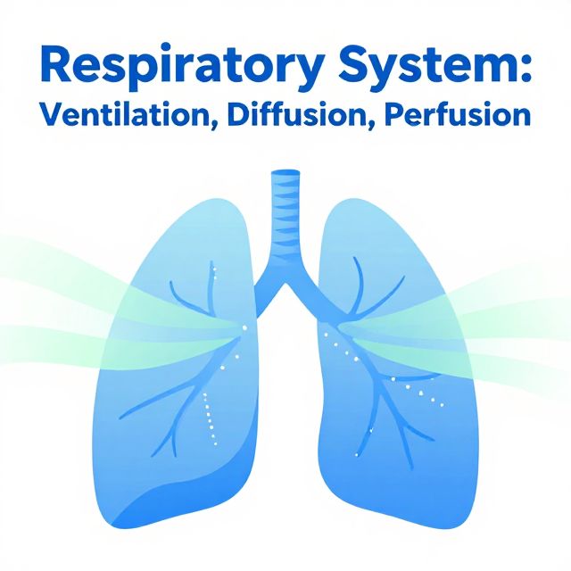
📚 Respiratory System: Fundamental Processes and Disorders
This study material compiles information from a lecture audio transcript and copy-pasted text, focusing on the core mechanisms of respiration and common associated disorders. It aims to provide a clear, organized overview for effective learning and review.
🌬️ Three Main Processes of Respiration
The human respiratory system relies on three interconnected processes to ensure efficient gas exchange: Ventilation, Perfusion, and Diffusion. Abnormalities in any of these can lead to significant health issues.
1. Ventilation
Ventilation is the mechanical process of moving air in and out of the lungs, facilitating gas exchange within the respiratory system.
- Pulmonary Ventilation: ✅ The total exchange of gases between the atmosphere and the lungs (breathing in and out).
- Alveolar Ventilation: ✅ The exchange of gases specifically within the gas-exchange portions of the lungs (alveoli).
Causes of Increased Ventilation: Increased ventilation can occur due to:
- Physiological reasons: Such as during physical exertion (work) when the body's oxygen demand increases.
- Pathophysiological reasons:
- Metabolic acidosis (body compensates by expelling more CO2).
- Increased demand for oxygen.
- Inappropriate hyperactivity of respiratory neurons.
Parameters of Ventilation: 📊
- Tidal Volume (VT): Volume of air moved during normal inspiration and expiration.
- Vital Capacity (VC): Maximum volume of air exhaled after a maximal inspiration.
- Maximal Breathing Capacity (V̇max): Maximal ventilation (L/min) achievable in a short period (usually 10 seconds).
- Compliance (C): Lung distensibility; how easily the lung can be stretched.
- Forced Expiration Volume in 1 second (FEV1): Maximal volume of air expired in the first second of a forced exhalation.
- Functional Residual Capacity (FRC): Total residual volume of air remaining in the lungs after a normal expiration.
2. Perfusion
Perfusion describes the continuous flow of blood through the pulmonary capillary bed, which is crucial for the gas exchange function of the lungs.
- Pulmonary Blood Vessels:
- Thinner and more compliant than systemic vessels.
- Offer less resistance to blood flow.
- Operate at much lower pressures (e.g., 22/8 mmHg) compared to systemic circulation (e.g., 120/70 mmHg).
3. Diffusion
Diffusion is the movement of gases (oxygen and carbon dioxide) in the alveoli and across the alveolar-capillary membrane.
Factors Influencing Diffusion: 💡
- Partial Pressure Difference: Higher concentration of oxygen increases the partial pressure difference across the membrane, enhancing diffusion. (e.g., oxygen therapy).
- Gas Characteristics: Molecular weight and solubility determine the diffusion coefficient.
- Carbon dioxide diffuses ~20 times faster than oxygen due to its greater solubility in respiratory membranes.
Factors Affecting Gas Exchange Surface Area:
- Surface area available for diffusion:
- Examples: Removal of a lung, emphysema, or chronic bronchitis (destroy lung tissue or cause V/Q mismatch).
- Thickness of the alveolar-capillary membrane:
- Examples: Pneumonia, interstitial lung disease, pulmonary edema (increase membrane thickness).
- Partial pressure of alveolar gases:
- Examples: High altitudes (reduced partial pressure of oxygen); oxygen therapy (increases gradient).
- Solubility and molecular weight of the gas:
- Examples: Carbon dioxide's high solubility allows it to diffuse much faster than oxygen.
⚠️ Consequences of Inadequate Breathing
Inadequate breathing can lead to critical conditions: Hypoxemia, Hypercapnia, and Hypocapnia.
1. Hypoxemia
Hypoxemia is a reduction in the partial pressure of oxygen (PO2) in the arterial blood.
Mechanisms of Hypoxemia:
- Hypoventilation: Inadequate air movement.
- Diffusion Impairment: Gases cannot effectively cross the alveolar-capillary membrane.
- Shunt: Blood bypasses the oxygenation process.
- Ventilation-Perfusion (V/Q) Impairment: Mismatch between air delivery and blood flow.
- Reduction of inspired oxygen partial pressure: Common at high altitudes.
Causes of Hypoventilation:
- Depression of the respiratory center (e.g., drug overdose).
- Diseases of nerves supplying respiratory muscles.
- Disorders of respiratory muscles (e.g., muscular dystrophy).
- Thoracic cage disorders (e.g., crushed chest).
- Decreased thoracic mobility (e.g., deformity, joint inflammation).
- Enlargement of the pleural space (e.g., pleural effusion, pneumothorax).
- Restrictive or obstructive lung disease.
Impaired Diffusion:
- Definition: Oxygen in the alveoli does not equilibrate with that in the pulmonary capillary blood.
- Causes:
- Altered thickness or permeability of the alveolar-capillary membrane.
- Increased distance from alveolar gas to red blood cells.
- Decreased permeability of the membrane to gas movement.
- Disorders: Interstitial lung disease, Adult Respiratory Distress Syndrome (ARDS), pulmonary edema, pneumonia.
Shunt: Blood moves from the right to the left side of circulation without being oxygenated.
- Physiologic Shunt: Mismatch of ventilation and perfusion; insufficient ventilation to oxygenate blood flowing through alveolar capillaries.
- Anatomic Shunt: Blood moves from venous to arterial side without passing through the lungs. Most are extrapulmonary (e.g., congenital heart disease). A completely unventilated lung portion (e.g., atelectasis) can also cause shunting.
Ventilation-Perfusion Mismatching: Occurs when lung areas are ventilated but not perfused, or perfused but not ventilated.
- Even normal lungs have some mismatch (e.g., top vs. bottom).
- In disease states (e.g., COPD), this relationship is severely disrupted, causing hypoxemia.
Manifestations of Hypoxemia:
- Arterial PO2 < 50 mmHg.
- Tachycardia, mild increase in blood pressure.
- Cool and moist skin.
- CNS effects: Confusion, delirium, difficulty in problem-solving, euphoria, stupor, coma (late).
- Late signs: Hypotension, bradycardia.
- Cyanosis: Bluish discoloration of skin/mucous membranes due to excessive deoxygenated hemoglobin (>5 g/dL). Most marked in lips, nail beds, ears, cheeks.
- Central Cyanosis: Increased deoxygenated hemoglobin in arterial blood; affects mucous membranes and skin.
- Peripheral Cyanosis: Slowing of blood flow to an area, increased oxygen extraction; results from vasoconstriction (e.g., cold exposure, shock, CHF).
2. Hypercapnia
Hypercapnia is defined as an arterial PCO2 > 44 mmHg.
Mechanisms of Hypercapnia:
- Alterations in carbon dioxide production.
- Abnormalities in respiratory function of chest wall and muscles.
- Changes in neural control of respiration.
Increased Carbon Dioxide Production:
- Changes in metabolic rate (activity, fever, disease).
- Example: CO2 production increases 13% for every 1°C increase in temperature above normal. Hypercapnia occurs if ventilation doesn't rise proportionally.
Disorders of Respiratory Muscle Function:
- Respiratory muscle fatigue (e.g., in primary respiratory diseases, neuromuscular disorders).
- Energy supply depends on blood flow and oxygen content. Low cardiac output, anemia, decreased oxygen saturation contribute to fatigue.
- Electrolyte imbalances (hypokalemia, hypophosphatemia) can cause muscle weakness.
Disorders of Neural Control of Respiration:
- Respiratory center (medulla oblongata and pons) activates respiratory muscles.
- CO2 crosses the blood-brain barrier easily.
- CO2 does not directly stimulate ventilation; it reacts with water to form carbonic acid, which dissociates into hydrogen ions (H+). H+ ions potently stimulate the respiratory center.
Manifestations of Hypercapnia:
- Affects respiratory, renal, neural, cardiovascular systems, and acid-base balance.
- Body adapts to chronic increases; symptoms may not appear until PCO2 > 50 mmHg.
- Symptoms:
- Headache.
- Hyperemic conjunctivae, flushed skin (due to CO2's vasodilatory effect).
- Carbon Dioxide Narcosis: Progressive somnolence, disorientation, coma (if untreated).
- PCO2 60-75 mmHg: Air hunger, rapid breathing.
- PCO2 80-100 mmHg: Lethargy.
- PCO2 100-150 mmHg: Anesthesia, death.
🗣️ Breathing Activities and Patterns
Terms for Various Breathing Activities 📚
- Eupnea: Normal breathing movements.
- Hyperpnea: Increased rate and depth of respiration.
- Hypopnea: Decreased breathing movements.
- Bradypnea: Decreased rate of breathing.
- Tachypnea: Increased rate of breathing.
- Apnea: Arrested breathing (cessation).
- Dyspnea: Subjective sensation of labored breathing.
- Asphyxia: Inability to breathe.
- Orthopnea: Labored breathing, relieved by sitting or upright position.
- Hyperventilation: Ventilation in excess of CO2 elimination needs, leading to decreased arterial PCO2 and respiratory alkalosis.
- Hypoventilation: Inadequate ventilation for gas exchange, leading to increased arterial PCO2, respiratory acidosis, and decreased arterial PO2.
Periodic Breathing Patterns
Describes patterns with episodes of apnea.
-
Cheyne-Stokes Breathing:
- Characterized by periods of slowly waxing and waning respirations separated by periods of apnea (up to 30 seconds).
- Mechanism: Impaired central feedback mechanisms buffering the respiratory center's response to CO2. During hyperpneic phase, CO2 falls, reducing stimulus and leading to apnea. Apnea causes CO2 accumulation, triggering next hyperpneic phase.
- Predisposing Conditions:
- Congestive heart failure (delay in blood transport to chemoreceptors).
- Impaired brain centers regulating respiration (e.g., brain lesions).
- Can occur in healthy persons at high altitudes (adaptive response, especially during sleep).
-
Biot's Breathing:
- Apnea recurs, but during ventilation, tidal volume and frequency remain fixed.
- Mechanism: Unclear, possibly a variant of Cheyne-Stokes.
- Associated Conditions: Central nervous system diseases, especially meningitis.
-
Kussmaul Respiration: Hyperventilation often seen in diabetes (diabetic ketoacidosis).
Dyspnea
- Definition: A subjective sensation of difficulty in breathing, including the perception of labored breathing and the reaction to that sensation.
- Associated Conditions:
- Primary lung diseases (pneumonia, asthma, emphysema).
- Heart disease with pulmonary congestion.
- Neuromuscular disorders affecting respiratory muscles (myasthenia gravis, muscular dystrophy).
- Proposed Mechanisms:
- Stimulation of lung receptors.
- Increased sensitivity to ventilation changes perceived via CNS.
- Reduced ventilatory capacity or breathing reserve.
- Stimulation of neural receptors in intercostal/diaphragm muscles and skeletal joints.
- Measurement: Often assessed by retrospective determination of activity level causing dyspnea (using scales).
- Treatment: Depends on the cause. Techniques include anxiety reduction, breathing retraining, and energy conservation measures.